5NPO | pdb_00005npo

Promiscuous Protein Self-Assembly as a Function of Protein Stability


Experimental Data Snapshot

  • Method: X-RAY DIFFRACTION
  • Resolution: 1.95 Å
  • R-Value Free: 
    0.256 (Depositor), 0.258 (DCC) 
  • R-Value Work: 
    0.199 (Depositor), 0.208 (DCC) 
  • R-Value Observed: 
    0.202 (Depositor) 

Starting Model: experimental
View more details

wwPDB Validation 3D Report Full Report

Validation slider image for 5NPO

This is version 1.2 of the entry. See complete history

Literature

Promiscuous Protein Binding as a Function of Protein Stability.

Cohen-Khait, R.Dym, O.Hamer-Rogotner, S.Schreiber, G.

(2017) Structure 25: 1867-1874.e3

  • DOI: https://doi.org/10.1016/j.str.2017.11.002
  • Primary Citation Related Structures: 
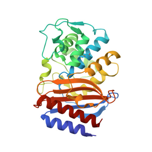
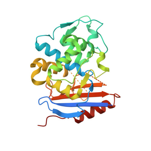
    5NPO

  • PubMed Abstract: 

    Proteins have evolved to balance efficient binding of desired partners with rejection of unwanted interactions. To investigate the evolution of protein-protein interactions, we selected a random library of pre-stabilized TEM1 β-lactamase against wild-type TEM1 using yeast surface display. Three mutations were sufficient to achieve micromolar affinity binding between the two. The X-ray structure emphasized that the main contribution of the selected mutations was to modify the protein fold, specifically removing the N'-terminal helix, which consequently allowed protein coupling via a β-sheet-mediated interaction resembling amyloid interaction mode. The only selected mutation located at the interaction interface (E58V) is reminiscent of the single mutation commonly causing sickle-cell anemia. Interestingly, the evolved mutations cannot be inserted into the wild-type protein due to reduced thermal stability of the resulting mutant protein. These results reveal a simple mechanism by which undesirable binding is purged by loss of thermal stability.


  • Organizational Affiliation
    • Department of Biomolecular Sciences, Weizmann Institute of Science, 76100 Rehovot, Israel.

Macromolecule Content 

  • Total Structure Weight: 58.07 kDa 
  • Atom Count: 3,744 
  • Modeled Residue Count: 486 
  • Deposited Residue Count: 529 
  • Unique protein chains: 2

Macromolecules

Find similar proteins by:|  3D Structure
Entity ID: 1
MoleculeChains  Sequence LengthOrganismDetailsImage
Beta-lactamase TEM264Escherichia coliMutation(s): 0 
Gene Names: blablaT-3blaT-4blaT-5blaT-6
EC: 3.5.2.6
UniProt
Find proteins for P62593 (Escherichia coli)
Explore P62593 
Go to UniProtKB:  P62593
Entity Groups
Sequence Clusters30% Identity50% Identity70% Identity90% Identity95% Identity100% Identity
UniProt GroupP62593
Sequence Annotations
Expand
Reference Sequence
Find similar proteins by:|  3D Structure
Entity ID: 2
MoleculeChains  Sequence LengthOrganismDetailsImage
Beta-lactamase265Escherichia coliMutation(s): 8 
EC: 3.5.2.6
UniProt
Find proteins for D3INY1 (Escherichia coli)
Explore D3INY1 
Go to UniProtKB:  D3INY1
Entity Groups
Sequence Clusters30% Identity50% Identity70% Identity90% Identity95% Identity100% Identity
UniProt GroupD3INY1
Sequence Annotations
Expand
Reference Sequence

Small Molecules

Ligands 1 Unique
IDChains Name / Formula / InChI Key2D Diagram3D Interactions
MG

Query on MG


Download:Ideal Coordinates CCD File
C [auth B]MAGNESIUM ION
Mg
JLVVSXFLKOJNIY-UHFFFAOYSA-N

Experimental Data & Validation

Experimental Data

  • Method: X-RAY DIFFRACTION
  • Resolution: 1.95 Å
  • R-Value Free:  0.256 (Depositor), 0.258 (DCC) 
  • R-Value Work:  0.199 (Depositor), 0.208 (DCC) 
  • R-Value Observed: 0.202 (Depositor) 
Space Group: P 1 21 1
Unit Cell:
Length ( Å )Angle ( ˚ )
a = 66.909α = 90
b = 52.733β = 90.59
c = 67.944γ = 90
Software Package:
Software NamePurpose
REFMACrefinement
XDSdata reduction
XDSdata scaling
PHASERphasing

Structure Validation

View Full Validation Report


Entry History 

Deposition Data

Revision History  (Full details and data files)

  • Version 1.0: 2017-12-20
    Type: Initial release
  • Version 1.1: 2024-01-17
    Changes: Data collection, Database references, Refinement description
  • Version 1.2: 2024-10-23
    Changes: Structure summary