5NKL | pdb_00005nkl

Crystal structure of the large fragment of DNA polymerase I from Thermus Aquaticus in a closed ternary complex with the artificial base pair dDs-dPxTP


Experimental Data Snapshot

  • Method: X-RAY DIFFRACTION
  • Resolution: 1.70 Å
  • R-Value Free: 
    0.237 (Depositor), 0.237 (DCC) 
  • R-Value Work: 
    0.181 (Depositor), 0.182 (DCC) 
  • R-Value Observed: 
    0.183 (Depositor) 

Starting Model: experimental
View more details

wwPDB Validation 3D Report Full Report

Validation slider image for 5NKL

Ligand Structure Quality Assessment 


This is version 1.3 of the entry. See complete history

Literature

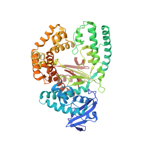
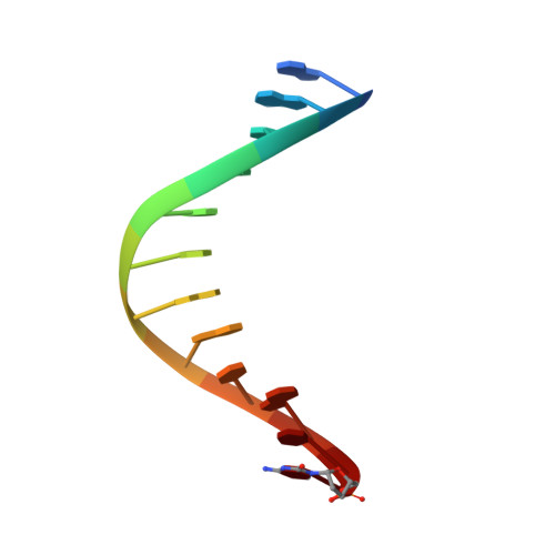
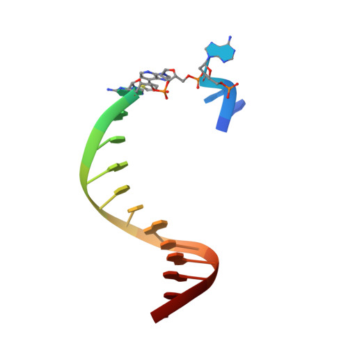
Structural Basis for Expansion of the Genetic Alphabet with an Artificial Nucleobase Pair.

Betz, K.Kimoto, M.Diederichs, K.Hirao, I.Marx, A.

(2017) Angew Chem Int Ed Engl 56: 12000-12003

  • DOI: https://doi.org/10.1002/anie.201704190
  • Primary Citation Related Structures: 
    5NKL

  • PubMed Abstract: 

    Hydrophobic artificial nucleobase pairs without the ability to pair through hydrogen bonds are promising candidates to expand the genetic alphabet. The most successful nucleobase surrogates show little similarity to each other and their natural counterparts. It is thus puzzling how these unnatural molecules are processed by DNA polymerases that have evolved to efficiently work with the natural building blocks. Here, we report structural insight into the insertion of one of the most promising hydrophobic unnatural base pairs, the dDs-dPx pair, into a DNA strand by a DNA polymerase. We solved a crystal structure of KlenTaq DNA polymerase with a modified template/primer duplex bound to the unnatural triphosphate. The ternary complex shows that the artificial pair adopts a planar structure just like a natural nucleobase pair, and identifies features that might hint at the mechanisms accounting for the lower incorporation efficiency observed when processing the unnatural substrates.


  • Organizational Affiliation
    • Departments of Chemistry and Biology and Konstanz Research School Chemical Biology, Universität Konstanz, Universitätsstrasse 10, 78464, Konstanz, Germany.

Macromolecule Content 

  • Total Structure Weight: 70.99 kDa 
  • Atom Count: 5,283 
  • Modeled Residue Count: 568 
  • Deposited Residue Count: 568 
  • Unique protein chains: 1
  • Unique nucleic acid chains: 2

Macromolecules


Find similar proteins by:|  3D Structure
Entity ID: 1
MoleculeChains  Sequence LengthOrganismDetailsImage
DNA polymerase I, thermostable540Thermus aquaticusMutation(s): 0 
Gene Names: polApol1
EC: 2.7.7.7
UniProt
Find proteins for P19821 (Thermus aquaticus)
Explore P19821 
Go to UniProtKB:  P19821
Entity Groups
Sequence Clusters30% Identity50% Identity70% Identity90% Identity95% Identity100% Identity
UniProt GroupP19821
Sequence Annotations
Expand
Reference Sequence
Find similar nucleic acids by:  Sequence
Entity ID: 2
MoleculeChains LengthOrganismImage
DNA (5'-D(*GP*AP*CP*CP*AP*CP*GP*GP*CP*GP*CP*(DOC))-3')12synthetic construct
Sequence Annotations
Expand
Reference Sequence
Find similar nucleic acids by:  Sequence
Entity ID: 3
MoleculeChains LengthOrganismImage
DNA (5'-D(*AP*AP*AP*(DNU)P*GP*GP*CP*GP*CP*CP*GP*TP*GP*GP*TP*C)-3')16synthetic construct
Sequence Annotations
Expand
Reference Sequence

Small Molecules

Ligands 5 Unique
IDChains Name / Formula / InChI Key2D Diagram3D Interactions
91T

Query on 91T


Download:Ideal Coordinates CCD File
G [auth A][[(2~{R},3~{S},5~{R})-5-[4-[(4~{R})-4,5-bis(oxidanyl)pent-1-ynyl]-2-nitro-pyrrol-1-yl]-3-oxidanyl-oxolan-2-yl]methoxy-oxidanyl-phosphoryl] phosphono hydrogen phosphate
C14 H21 N2 O16 P3
JIVPAQSXWIKFCI-GFQSEFKGSA-N
91R

Query on 91R


Download:Ideal Coordinates CCD File
H [auth A][[(2~{R},3~{S},5~{R})-5-[4-[(4~{S})-4,5-bis(oxidanyl)pent-1-ynyl]-2-nitro-pyrrol-1-yl]-3-oxidanyl-oxolan-2-yl]methoxy-oxidanyl-phosphoryl] phosphono hydrogen phosphate
C14 H21 N2 O16 P3
JIVPAQSXWIKFCI-CIQGVGRVSA-N
GOL

Query on GOL


Download:Ideal Coordinates CCD File
I [auth C],
J [auth C]
GLYCEROL
C3 H8 O3
PEDCQBHIVMGVHV-UHFFFAOYSA-N
ACT

Query on ACT


Download:Ideal Coordinates CCD File
F [auth A]ACETATE ION
C2 H3 O2
QTBSBXVTEAMEQO-UHFFFAOYSA-M
MG

Query on MG


Download:Ideal Coordinates CCD File
D [auth A],
E [auth A]
MAGNESIUM ION
Mg
JLVVSXFLKOJNIY-UHFFFAOYSA-N

Experimental Data & Validation

Experimental Data

  • Method: X-RAY DIFFRACTION
  • Resolution: 1.70 Å
  • R-Value Free:  0.237 (Depositor), 0.237 (DCC) 
  • R-Value Work:  0.181 (Depositor), 0.182 (DCC) 
  • R-Value Observed: 0.183 (Depositor) 
Space Group: P 31 2 1
Unit Cell:
Length ( Å )Angle ( ˚ )
a = 108.777α = 90
b = 108.777β = 90
c = 90.571γ = 120
Software Package:
Software NamePurpose
PHENIXrefinement
XDSdata reduction
XDSdata scaling
PHENIXphasing

Structure Validation

View Full Validation Report


Ligand Structure Quality Assessment 


Entry History 

Deposition Data

Revision History  (Full details and data files)

  • Version 1.0: 2017-06-28
    Type: Initial release
  • Version 1.1: 2017-07-05
    Changes: Database references
  • Version 1.2: 2017-09-27
    Changes: Database references
  • Version 1.3: 2024-01-17
    Changes: Data collection, Database references, Derived calculations, Refinement description