


Crystal structure of the methyltransferase subunit of human mitochondrial Ribonuclease P (MRPP1) bound to S-adenosyl-methionine (SAM)
Oerum, S., Kopec, J., Fitzpatrick, F., Newman, J.A., Oppermann, U., Yue, W.W.To be published.
Experimental Data Snapshot
Entity ID: 1 | |||||
|---|---|---|---|---|---|
| Molecule | Chains | Sequence Length | Organism | Details | Image |
| Mitochondrial ribonuclease P protein 1 | 202 | Homo sapiens | Mutation(s): 0 Gene Names: TRMT10C, MRPP1, RG9MTD1 EC: 2.1.1 (PDB Primary Data), 2.1.1.221 (UniProt), 2.1.1.218 (UniProt) | 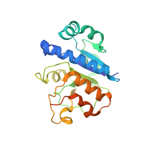 | |
UniProt & NIH Common Fund Data Resources | |||||
PHAROS: Q7L0Y3 GTEx: ENSG00000174173 | |||||
Entity Groups | |||||
| Sequence Clusters | 30% Identity50% Identity70% Identity90% Identity95% Identity100% Identity | ||||
| UniProt Group | Q7L0Y3 | ||||
Sequence AnnotationsExpand | |||||
Reference Sequence | |||||
| Ligands 3 Unique | |||||
|---|---|---|---|---|---|
| ID | Chains | Name / Formula / InChI Key | 2D Diagram | 3D Interactions | |
| SAM Download:Ideal Coordinates CCD File | D [auth A], J [auth B], P [auth C] | S-ADENOSYLMETHIONINE C15 H22 N6 O5 S MEFKEPWMEQBLKI-FCKMPRQPSA-N |  | ||
| GOL Download:Ideal Coordinates CCD File | E [auth A] F [auth A] G [auth A] K [auth B] L [auth B] | GLYCEROL C3 H8 O3 PEDCQBHIVMGVHV-UHFFFAOYSA-N |  | ||
| EDO Download:Ideal Coordinates CCD File | H [auth A], I [auth A], N [auth B], O [auth B] | 1,2-ETHANEDIOL C2 H6 O2 LYCAIKOWRPUZTN-UHFFFAOYSA-N |  | ||
| Length ( Å ) | Angle ( ˚ ) |
|---|---|
| a = 82.64 | α = 90 |
| b = 82.64 | β = 90 |
| c = 148.62 | γ = 90 |
| Software Name | Purpose |
|---|---|
| REFMAC | refinement |
| XDS | data reduction |
| Aimless | data scaling |
| SHELXD | phasing |


