5NB7 | pdb_00005nb7

Complement factor D


Experimental Data Snapshot

  • Method: X-RAY DIFFRACTION
  • Resolution: 1.33 Å
  • R-Value Free: 
    0.196 (Depositor), 0.202 (DCC) 
  • R-Value Work: 
    0.140 (Depositor), 0.148 (DCC) 
  • R-Value Observed: 
    0.143 (Depositor) 

Starting Model: experimental
View more details

wwPDB Validation 3D Report Full Report

Validation slider image for 5NB7

Ligand Structure Quality Assessment 


This is version 1.3 of the entry. See complete history

Literature

Discovery of Highly Potent and Selective Small-Molecule Reversible Factor D Inhibitors Demonstrating Alternative Complement Pathway Inhibition in Vivo.

Lorthiois, E.Anderson, K.Vulpetti, A.Rogel, O.Cumin, F.Ostermann, N.Steinbacher, S.Mac Sweeney, A.Delgado, O.Liao, S.M.Randl, S.Rudisser, S.Dussauge, S.Fettis, K.Kieffer, L.de Erkenez, A.Yang, L.Hartwieg, C.Argikar, U.A.La Bonte, L.R.Newton, R.Kansara, V.Flohr, S.Hommel, U.Jaffee, B.Maibaum, J.

(2017) J Med Chem 60: 5717-5735

  • DOI: https://doi.org/10.1021/acs.jmedchem.7b00425
  • Primary Citation Related Structures: 
    5NAR, 5NAT, 5NAW, 5NB6, 5NB7, 5NBA

  • PubMed Abstract: 

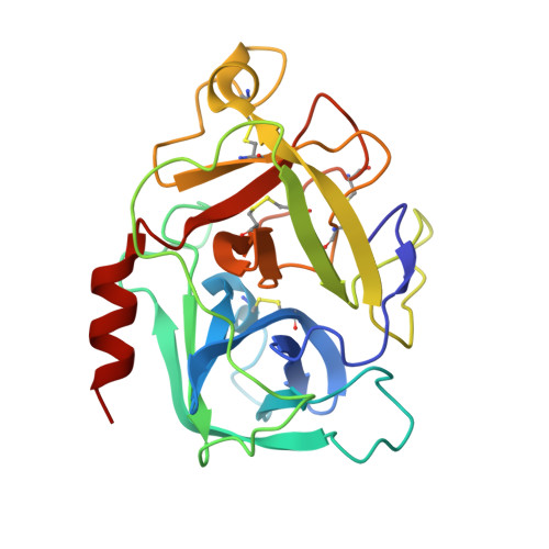
    The highly specific S1 serine protease factor D (FD) plays a central role in the amplification of the complement alternative pathway (AP) of the innate immune system. Genetic associations in humans have implicated AP activation in age-related macular degeneration (AMD), and AP dysfunction predisposes individuals to disorders such as paroxysmal nocturnal hemoglobinuria (PNH) and atypical hemolytic uremic syndrome (aHUS). The combination of structure-based hit identification and subsequent optimization of the center (S)-proline-based lead 7 has led to the discovery of noncovalent reversible and selective human factor D (FD) inhibitors with drug-like properties. The orally bioavailable compound 2 exerted excellent potency in 50% human whole blood in vitro and blocked AP activity ex vivo after oral administration to monkeys as demonstrated by inhibition of membrane attack complex (MAC) formation. Inhibitor 2 demonstrated sustained oral and ocular efficacy in a model of lipopolysaccharide (LPS)-induced systemic AP activation in mice expressing human FD.


  • Organizational Affiliation
    • Novartis Pharma AG, Novartis Institutes for BioMedical Research , Novartis Campus, CH-4056 Basel, Switzerland.

Macromolecule Content 

  • Total Structure Weight: 25.24 kDa 
  • Atom Count: 1,961 
  • Modeled Residue Count: 230 
  • Deposited Residue Count: 230 
  • Unique protein chains: 1

Macromolecules

Find similar proteins by:|  3D Structure
Entity ID: 1
MoleculeChains  Sequence LengthOrganismDetailsImage
Complement factor D230Homo sapiensMutation(s): 0 
Gene Names: CFDDFPFD
EC: 3.4.21.46
UniProt & NIH Common Fund Data Resources
Find proteins for P00746 (Homo sapiens)
Explore P00746 
Go to UniProtKB:  P00746
PHAROS:  P00746
GTEx:  ENSG00000197766 
Entity Groups
Sequence Clusters30% Identity50% Identity70% Identity90% Identity95% Identity100% Identity
UniProt GroupP00746
Sequence Annotations
Expand
Reference Sequence

Small Molecules

Ligands 2 Unique
IDChains Name / Formula / InChI Key2D Diagram3D Interactions
8NQ

Query on 8NQ


Download:Ideal Coordinates CCD File
D [auth A]1-[2-[(1~{R},3~{S},5~{R})-3-[(6-bromanylpyridin-2-yl)carbamoyl]-2-azabicyclo[3.1.0]hexan-2-yl]-2-oxidanylidene-ethyl]indazole-3-carboxamide
C21 H19 Br N6 O3
CWZUTHDJLNZLCM-DFBGVHRSSA-N
DMS

Query on DMS


Download:Ideal Coordinates CCD File
B [auth A],
C [auth A]
DIMETHYL SULFOXIDE
C2 H6 O S
IAZDPXIOMUYVGZ-UHFFFAOYSA-N
Binding Affinity Annotations 
IDSourceBinding Affinity
8NQ BindingDB:  5NB7 IC50: min: 6, max: 20 (nM) from 4 assay(s)

Experimental Data & Validation

Experimental Data

  • Method: X-RAY DIFFRACTION
  • Resolution: 1.33 Å
  • R-Value Free:  0.196 (Depositor), 0.202 (DCC) 
  • R-Value Work:  0.140 (Depositor), 0.148 (DCC) 
  • R-Value Observed: 0.143 (Depositor) 
Space Group: P 1 21 1
Unit Cell:
Length ( Å )Angle ( ˚ )
a = 55.801α = 90
b = 49.884β = 106.08
c = 39.088γ = 90
Software Package:
Software NamePurpose
REFMACrefinement
XDSdata reduction
XSCALEdata scaling
PHASERphasing

Structure Validation

View Full Validation Report


Ligand Structure Quality Assessment 


Entry History 

Deposition Data

Revision History  (Full details and data files)

  • Version 1.0: 2017-06-28
    Type: Initial release
  • Version 1.1: 2017-07-26
    Changes: Database references
  • Version 1.2: 2024-01-17
    Changes: Data collection, Database references, Refinement description
  • Version 1.3: 2024-11-06
    Changes: Structure summary