5NB3

High resolution C-phycoerythrin from marine cyanobacterium Phormidium sp. A09DM at pH 7.5


Experimental Data Snapshot

  • Method: X-RAY DIFFRACTION
  • Resolution: 1.38 Å
  • R-Value Free: 0.211 
  • R-Value Work: 0.155 
  • R-Value Observed: 0.158 

wwPDB Validation   3D Report Full Report


Ligand Structure Quality Assessment 


This is version 1.3 of the entry. See complete history


Literature

An improved crystal structure of C-phycoerythrin from the marine cyanobacterium Phormidium sp. A09DM.

Sonani, R.R.Roszak, A.W.Ortmann de Percin Northumberland, C.Madamwar, D.Cogdell, R.J.

(2018) Photosynth Res 135: 65-78

  • DOI: https://doi.org/10.1007/s11120-017-0443-2
  • Primary Citation of Related Structures:  
    5NB3, 5NB4

  • PubMed Abstract: 

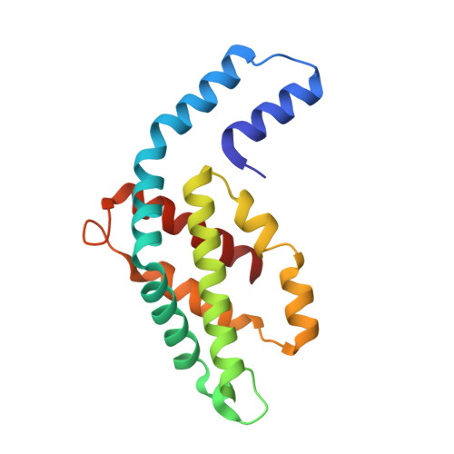
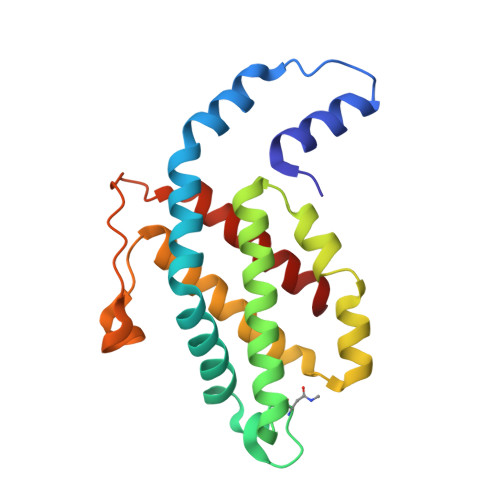
    C-Phycoerythrin (PE) from Phormidium sp. A09DM has been crystallized using different conditions and its structure determined to atomic resolution (1.14 Å). In order for the pigment present, phycoerythrobilin (PEB), to function as an efficient light-harvesting molecule it must be held rigidly (Kupka and Scheer in Biochim Biophys Acta 1777:94-103, 2008) and, moreover, the different PEB molecules in PE must be arranged, relative to each other, so as to promote efficient energy transfer between them. This improved structure has allowed us to define in great detail the structure of the PEBs and their binding sites. These precise structural details will facilitate theoretical calculations of each PEB's spectroscopic properties. It was possible, however, to suggest a model for which chromophores contribute to the different regions of absorption spectrum and propose a tentative scheme for energy transfer. We show that some subtle differences in one of these PEB binding sites in two of the 12 subunits are caused by crystal contacts between neighboring hexamers in the crystal lattice. This explains some of the differences seen in previous lower resolution structures determined at two different pH values (Kumar et al. in Photosyn Res 129:17-28, 2016).


  • Organizational Affiliation

    Post-Graduate Department of Biosciences, UGC-Centre of Advanced Study, Sardar Patel University, Vadtal Road, Satellite Campus, Bakrol, Anand, Gujarat, 388315, India.


Macromolecules
Find similar proteins by:  (by identity cutoff)  |  3D Structure
Entity ID: 1
MoleculeChains Sequence LengthOrganismDetailsImage
Phycoerythrin Alpha subunit,Phycoerythrin Alpha subunit,Phycoerythrin Alpha subunit
A, B, C, D, E
A, B, C, D, E, F, G, H, I, J, K, L
164Phormidium rubidum A09DMMutation(s): 0 
UniProt
Find proteins for A0A0E3W010 (Phormidium rubidum A09DM)
Explore A0A0E3W010 
Go to UniProtKB:  A0A0E3W010
Entity Groups  
Sequence Clusters30% Identity50% Identity70% Identity90% Identity95% Identity100% Identity
UniProt GroupA0A0E3W010
Sequence Annotations
Expand
  • Reference Sequence
Find similar proteins by:  (by identity cutoff)  |  3D Structure
Entity ID: 2
MoleculeChains Sequence LengthOrganismDetailsImage
Phycoerythrin Beta subunit,Phycoerythrin Beta subunit
M, N, O, P, Q
M, N, O, P, Q, R, S, T, U, V, W, X
184Phormidium rubidum A09DMMutation(s): 0 
UniProt
Find proteins for A0A0E4G455 (Phormidium rubidum A09DM)
Explore A0A0E4G455 
Go to UniProtKB:  A0A0E4G455
Entity Groups  
Sequence Clusters30% Identity50% Identity70% Identity90% Identity95% Identity100% Identity
UniProt GroupA0A0E4G455
Sequence Annotations
Expand
  • Reference Sequence
Small Molecules
Ligands 6 Unique
IDChains Name / Formula / InChI Key2D Diagram3D Interactions
PEB
Query on PEB

Download Ideal Coordinates CCD File 
AB [auth H]
AC [auth N]
AD [auth R]
AE [auth W]
BB [auth H]
AB [auth H],
AC [auth N],
AD [auth R],
AE [auth W],
BB [auth H],
BC [auth N],
CC [auth N],
DD [auth S],
EA [auth B],
EB [auth I],
ED [auth S],
EE [auth X],
FA [auth B],
FB [auth I],
FD [auth S],
FE [auth X],
GE [auth X],
HC [auth O],
HD [auth T],
IA [auth C],
IB [auth J],
IC [auth O],
ID [auth T],
JA [auth C],
JB [auth J],
JC [auth O],
JD [auth T],
LD [auth U],
MC [auth P],
MD [auth U],
NA [auth D],
NC [auth P],
ND [auth U],
OA [auth D],
OB [auth K],
OC [auth P],
PB [auth K],
RA [auth E],
RD [auth V],
SA [auth E],
SB [auth L],
SC [auth Q],
SD [auth V],
TB [auth L],
TC [auth Q],
TD [auth V],
UA [auth F],
UC [auth Q],
VA [auth F],
WB [auth M],
XA [auth G],
XB [auth M],
Y [auth A],
YA [auth G],
YB [auth M],
YC [auth R],
YD [auth W],
Z [auth A],
ZC [auth R],
ZD [auth W]
PHYCOERYTHROBILIN
C33 H40 N4 O6
NKCBCVIFPXGHAV-WAVSMFBNSA-N
MRD
Query on MRD

Download Ideal Coordinates CCD File 
KD [auth T],
UD [auth V]
(4R)-2-METHYLPENTANE-2,4-DIOL
C6 H14 O2
SVTBMSDMJJWYQN-RXMQYKEDSA-N
MPD
Query on MPD

Download Ideal Coordinates CCD File 
BD [auth R]
BE [auth W]
CE [auth W]
DC [auth N]
EC [auth N]
BD [auth R],
BE [auth W],
CE [auth W],
DC [auth N],
EC [auth N],
GD [auth S],
HE [auth X],
KC [auth O],
OD [auth U],
PC [auth P],
VC [auth Q],
ZB [auth M]
(4S)-2-METHYL-2,4-PENTANEDIOL
C6 H14 O2
SVTBMSDMJJWYQN-YFKPBYRVSA-N
FMT
Query on FMT

Download Ideal Coordinates CCD File 
AA [auth A]
BA [auth A]
CB [auth H]
DE [auth W]
FC [auth N]
AA [auth A],
BA [auth A],
CB [auth H],
DE [auth W],
FC [auth N],
KA [auth C],
KB [auth J],
LC [auth O],
PD [auth U],
QB [auth K],
QC [auth P],
QD [auth U],
VD [auth V],
WC [auth Q],
WD [auth V]
FORMIC ACID
C H2 O2
BDAGIHXWWSANSR-UHFFFAOYSA-N
CL
Query on CL

Download Ideal Coordinates CCD File 
DA [auth A]
HA [auth B]
HB [auth I]
MA [auth C]
NB [auth J]
DA [auth A],
HA [auth B],
HB [auth I],
MA [auth C],
NB [auth J],
VB [auth L]
CHLORIDE ION
Cl
VEXZGXHMUGYJMC-UHFFFAOYSA-M
NA
Query on NA

Download Ideal Coordinates CCD File 
CA [auth A]
CD [auth R]
DB [auth H]
GA [auth B]
GB [auth I]
CA [auth A],
CD [auth R],
DB [auth H],
GA [auth B],
GB [auth I],
GC [auth N],
LA [auth C],
LB [auth J],
MB [auth J],
PA [auth D],
QA [auth D],
RB [auth K],
RC [auth P],
TA [auth E],
UB [auth L],
WA [auth F],
XC [auth Q],
XD [auth V],
ZA [auth G]
SODIUM ION
Na
FKNQFGJONOIPTF-UHFFFAOYSA-N
Modified Residues  1 Unique
IDChains TypeFormula2D DiagramParent
MEN
Query on MEN
M, N, O, P, Q
M, N, O, P, Q, R, S, T, U, V, W, X
L-PEPTIDE LINKINGC5 H10 N2 O3ASN
Experimental Data & Validation

Experimental Data

  • Method: X-RAY DIFFRACTION
  • Resolution: 1.38 Å
  • R-Value Free: 0.211 
  • R-Value Work: 0.155 
  • R-Value Observed: 0.158 
  • Space Group: P 1
Unit Cell:
Length ( Å )Angle ( ˚ )
a = 110.046α = 78.76
b = 110.17β = 82.28
c = 118.516γ = 60.43
Software Package:
Software NamePurpose
REFMACrefinement
XDSdata reduction
Aimlessdata scaling
REFMACphasing

Structure Validation

View Full Validation Report


Ligand Structure Quality Assessment 


Entry History & Funding Information

Deposition Data


Funding OrganizationLocationGrant Number
Department of Energy (DOE, United States)United StatesDE-SC0001035

Revision History  (Full details and data files)

  • Version 1.0: 2017-09-27
    Type: Initial release
  • Version 1.1: 2018-02-07
    Changes: Database references
  • Version 1.2: 2022-03-30
    Changes: Author supporting evidence, Database references, Derived calculations
  • Version 1.3: 2024-01-17
    Changes: Data collection, Refinement description