5N8S

Crystal Structure of Drosophila DHX36 helicase in complex with polyT


Experimental Data Snapshot

  • Method: X-RAY DIFFRACTION
  • Resolution: 2.88 Å
  • R-Value Free: 0.285 
  • R-Value Work: 0.207 
  • R-Value Observed: 0.211 

wwPDB Validation   3D Report Full Report


This is version 1.2 of the entry. See complete history


Literature

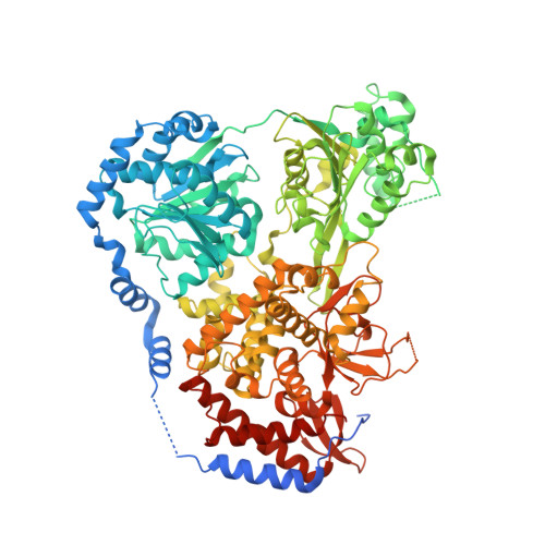
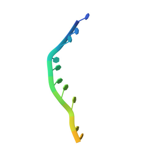
Molecular Mechanistic Insights into Drosophila DHX36-Mediated G-Quadruplex Unfolding: A Structure-Based Model.

Chen, W.F.Rety, S.Guo, H.L.Dai, Y.X.Wu, W.Q.Liu, N.N.Auguin, D.Liu, Q.W.Hou, X.M.Dou, S.X.Xi, X.G.

(2018) Structure 26: 403-415.e4

  • DOI: https://doi.org/10.1016/j.str.2018.01.008
  • Primary Citation of Related Structures:  
    5N8R, 5N8S, 5N90, 5N94, 5N96, 5N98, 5N9A, 5N9D

  • PubMed Abstract: 

    Helicase DHX36 plays essential roles in cell development and differentiation at least partially by resolving G-quadruplex (G4) structures. Here we report crystal structures of the Drosophila homolog of DHX36 (DmDHX36) in complex with RNA and a series of DNAs. By combining structural, small-angle X-ray scattering, molecular dynamics simulation, and single-molecule fluorescence studies, we revealed that positively charged amino acids in RecA2 and OB-like domains constitute an elaborate structural pocket at the nucleic acid entrance, in which negatively charged G4 DNA is tightly bound and partially destabilized. The G4 DNA is then completely unfolded through the 3'-5' translocation activity of the helicase. Furthermore, crystal structures and DNA binding assays show that G-rich DNA is preferentially recognized and in the presence of ATP, specifically bound by DmDHX36, which may cooperatively enhance the G-rich DNA translocation and G4 unfolding. On the basis of these results, a conceptual G4 DNA-resolving mechanism is proposed.


  • Organizational Affiliation

    College of Life Sciences, Northwest A&F University, Yangling, Shaanxi 712100, China.


Macromolecules

Find similar proteins by:  (by identity cutoff)  |  3D Structure
Entity ID: 1
MoleculeChains Sequence LengthOrganismDetailsImage
CG9323, isoform A
A, B
944Drosophila melanogasterMutation(s): 0 
Gene Names: CG9323Dmel_CG9323
EC: 3.6.1.3
UniProt
Find proteins for Q8SWT2 (Drosophila melanogaster)
Explore Q8SWT2 
Go to UniProtKB:  Q8SWT2
Entity Groups  
Sequence Clusters30% Identity50% Identity70% Identity90% Identity95% Identity100% Identity
UniProt GroupQ8SWT2
Sequence Annotations
Expand
  • Reference Sequence

Find similar nucleic acids by:  Sequence   |   3D Structure  

Entity ID: 2
MoleculeChains LengthOrganismImage
DNA (5'-D(P*TP*TP*TP*TP*TP*TP*TP*TP*TP*T)-3')
C, D
16Homo sapiens
Sequence Annotations
Expand
  • Reference Sequence
Experimental Data & Validation

Experimental Data

  • Method: X-RAY DIFFRACTION
  • Resolution: 2.88 Å
  • R-Value Free: 0.285 
  • R-Value Work: 0.207 
  • R-Value Observed: 0.211 
  • Space Group: C 1 2 1
Unit Cell:
Length ( Å )Angle ( ˚ )
a = 297.981α = 90
b = 49.923β = 115.01
c = 163.847γ = 90
Software Package:
Software NamePurpose
PHENIXrefinement
XDSdata reduction
Aimlessdata scaling
PHASERphasing

Structure Validation

View Full Validation Report


Entry History 

Deposition Data

Revision History  (Full details and data files)

  • Version 1.0: 2018-03-14
    Type: Initial release
  • Version 1.1: 2019-02-20
    Changes: Advisory, Data collection, Derived calculations
  • Version 1.2: 2024-01-17
    Changes: Data collection, Database references, Refinement description