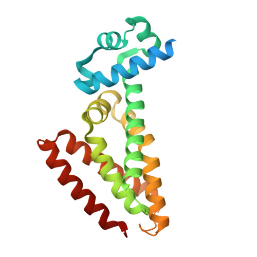
5N7O

EthR2 in complex with SMARt-420 compound


Experimental Data Snapshot

  • Method: X-RAY DIFFRACTION
  • Resolution: 2.30 Å
  • R-Value Free: 0.248 
  • R-Value Work: 0.215 
  • R-Value Observed: 0.217 

wwPDB Validation   3D Report Full Report


Ligand Structure Quality Assessment 


This is version 1.2 of the entry. See complete history


Literature

Structural analysis of the interaction between spiroisoxazoline SMARt-420 and the Mycobacterium tuberculosis repressor EthR2.

Wohlkonig, A.Remaut, H.Moune, M.Tanina, A.Meyer, F.Desroses, M.Steyaert, J.Willand, N.Baulard, A.R.Wintjens, R.

(2017) Biochem Biophys Res Commun 487: 403-408

  • DOI: https://doi.org/10.1016/j.bbrc.2017.04.074
  • Primary Citation of Related Structures:  
    5N7O

  • PubMed Abstract: 

    Inhibition of transcriptional regulators of bacterial pathogens with the aim of reprogramming their metabolism to modify their antibiotic susceptibility constitutes a promising therapeutic strategy. One example is the bio-activation of the anti-tubercular pro-drug ethionamide, which activity could be enhanced by inhibiting the transcriptional repressor EthR. Recently, we discovered that inhibition of a second transcriptional repressor, EthR2, leads to the awakening of a new ethionamide bio-activation pathway. The x-ray structure of EthR2 was solved at 2.3 Å resolution in complex with a compound called SMARt-420 (Small Molecule Aborting Resistance). Detailed comparison and structural analysis revealed interesting insights for the upcoming structure-based design of EthR2 inhibitors as an alternative to revert ethionamide resistance in Mycobacterium tuberculosis.


  • Organizational Affiliation

    Center for Structural Biology, Vlaams Instituut voor Biotechnology (VIB), Pleinlaan 2, B-1050 Brussels, Belgium; Structural Biology Brussels, Vrije Universiteit Brussel (VUB), Pleinlaan 2, B-1050 Brussels, Belgium.


Macromolecules
Find similar proteins by:  (by identity cutoff)  |  3D Structure
Entity ID: 1
MoleculeChains Sequence LengthOrganismDetailsImage
Probable transcriptional regulatory protein
A, B
221Mycobacterium tuberculosis H37RvMutation(s): 0 
Gene Names: Rv0078LH57_00450
UniProt
Find proteins for O53623 (Mycobacterium tuberculosis (strain ATCC 25618 / H37Rv))
Explore O53623 
Go to UniProtKB:  O53623
Entity Groups  
Sequence Clusters30% Identity50% Identity70% Identity90% Identity95% Identity100% Identity
UniProt GroupO53623
Sequence Annotations
Expand
  • Reference Sequence
Small Molecules
Ligands 1 Unique
IDChains Name / Formula / InChI Key2D Diagram3D Interactions
69Y
Query on 69Y

Download Ideal Coordinates CCD File 
C [auth A],
D [auth B]
4,4,4-trifluoro-1-(3-phenyl-1-oxa-2,8-diazaspiro[4.5]dec-2-en-8-yl)butan-1-one
C17 H19 F3 N2 O2
IBVDSAFESRHIBV-UHFFFAOYSA-N
Experimental Data & Validation

Experimental Data

  • Method: X-RAY DIFFRACTION
  • Resolution: 2.30 Å
  • R-Value Free: 0.248 
  • R-Value Work: 0.215 
  • R-Value Observed: 0.217 
  • Space Group: P 21 21 21
Unit Cell:
Length ( Å )Angle ( ˚ )
a = 59.4α = 90
b = 74.29β = 90
c = 91.5γ = 90
Software Package:
Software NamePurpose
REFMACrefinement
XDSdata reduction
XDSdata scaling
MOLREPphasing

Structure Validation

View Full Validation Report


Ligand Structure Quality Assessment 


Entry History 

Deposition Data

Revision History  (Full details and data files)

  • Version 1.0: 2017-04-26
    Type: Initial release
  • Version 1.1: 2017-05-10
    Changes: Database references
  • Version 1.2: 2024-01-17
    Changes: Data collection, Database references, Refinement description