5N7D

MAGI-1 complexed with a RSK1 peptide


Experimental Data Snapshot

  • Method: X-RAY DIFFRACTION
  • Resolution: 2.30 Å
  • R-Value Free: 0.227 
  • R-Value Work: 0.191 
  • R-Value Observed: 0.192 

wwPDB Validation   3D Report Full Report


This is version 2.0 of the entry. See complete history


Literature

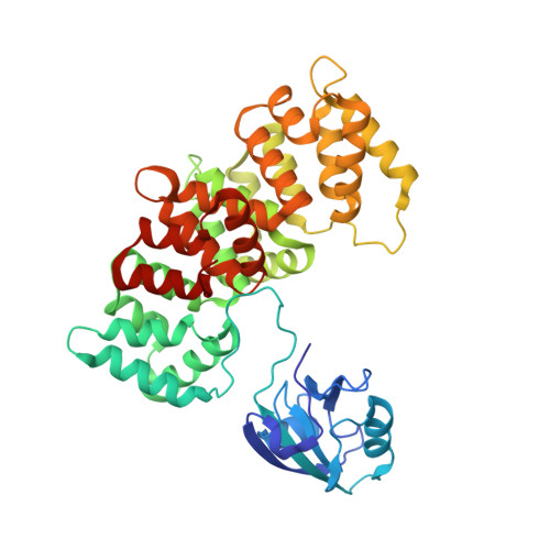
Dynamic control of RSK complexes by phosphoswitch-based regulation.

Gogl, G.Biri-Kovacs, B.Poti, A.L.Vadaszi, H.Szeder, B.Bodor, A.Schlosser, G.Acs, A.Turiak, L.Buday, L.Alexa, A.Nyitray, L.Remenyi, A.

(2018) FEBS J 285: 46-71

  • DOI: https://doi.org/10.1111/febs.14311
  • Primary Citation of Related Structures:  
    5N7D, 5N7F, 5N7G

  • PubMed Abstract: 

    Assembly and disassembly of protein-protein complexes needs to be dynamically controlled and phosphoswitches based on linear motifs are crucial in this process. Extracellular signal-regulated kinase 2 (ERK2) recognizes a linear-binding motif at the C-terminal tail (CTT) of ribosomal S6 kinase 1 (RSK1), leading to phosphorylation and subsequent activation of RSK1. The CTT also contains a classical PDZ domain-binding motif which binds RSK substrates (e.g. MAGI-1). We show that autophosphorylation of the disordered CTT promotes the formation of an intramolecular charge clamp, which efficiently masks critical residues and indirectly hinders ERK binding. Thus, RSK1 CTT operates as an autoregulated phosphoswitch: its phosphorylation at specific sites affects its protein-binding capacity and its conformational dynamics. These biochemical feedbacks, which form the structural basis for the rapid dissociation of ERK2-RSK1 and RSK1-PDZ substrate complexes under sustained epidermal growth factor (EGF) stimulation, were structurally characterized and validated in living cells. Overall, conformational changes induced by phosphorylation in disordered regions of protein kinases, coupled to allosteric events occurring in the kinase domain cores, may provide mechanisms that contribute to the emergence of complex signaling activities. In addition, we show that phosphoswitches based on linear motifs can be functionally classified as ON and OFF protein-protein interaction switches or dimmers, depending on the specific positioning of phosphorylation target sites in relation to functional linear-binding motifs. Moreover, interaction of phosphorylated residues with positively charged residues in disordered regions is likely to be a common mechanism of phosphoregulation.


  • Organizational Affiliation

    Department of Biochemistry, ELTE Eötvös Loránd University, Budapest, Hungary.


Macromolecules
Find similar proteins by:  (by identity cutoff)  |  3D Structure
Entity ID: 1
MoleculeChains Sequence LengthOrganismDetailsImage
Membrane-associated guanylate kinase, WW and PDZ domain-containing protein 1,Annexin A2
A, B
427Homo sapiensMutation(s): 0 
Gene Names: MAGI1AIP3BAIAP1BAP1TNRC19ANXA2ANX2ANX2L4CAL1HLPC2D
UniProt & NIH Common Fund Data Resources
Find proteins for Q96QZ7 (Homo sapiens)
Explore Q96QZ7 
Go to UniProtKB:  Q96QZ7
PHAROS:  Q96QZ7
GTEx:  ENSG00000151276 
Find proteins for P07355 (Homo sapiens)
Explore P07355 
Go to UniProtKB:  P07355
PHAROS:  P07355
GTEx:  ENSG00000182718 
Entity Groups  
Sequence Clusters30% Identity50% Identity70% Identity90% Identity95% Identity100% Identity
UniProt GroupsQ96QZ7P07355
Sequence Annotations
Expand
  • Reference Sequence
Find similar proteins by:  (by identity cutoff)  |  3D Structure
Entity ID: 2
MoleculeChains Sequence LengthOrganismDetailsImage
Ribosomal protein S6 kinase alpha-149Homo sapiensMutation(s): 0 
Gene Names: RPS6KA1MAPKAPK1ARSK1
EC: 2.7.11.1
UniProt & NIH Common Fund Data Resources
Find proteins for Q15418 (Homo sapiens)
Explore Q15418 
Go to UniProtKB:  Q15418
PHAROS:  Q15418
GTEx:  ENSG00000117676 
Entity Groups  
Sequence Clusters30% Identity50% Identity70% Identity90% Identity95% Identity100% Identity
UniProt GroupQ15418
Sequence Annotations
Expand
  • Reference Sequence
Small Molecules
Ligands 2 Unique
IDChains Name / Formula / InChI Key2D Diagram3D Interactions
GOL
Query on GOL

Download Ideal Coordinates CCD File 
D [auth A],
E [auth A],
F [auth A],
K [auth B],
L [auth B]
GLYCEROL
C3 H8 O3
PEDCQBHIVMGVHV-UHFFFAOYSA-N
CA
Query on CA

Download Ideal Coordinates CCD File 
G [auth A]
H [auth A]
I [auth A]
J [auth A]
M [auth B]
G [auth A],
H [auth A],
I [auth A],
J [auth A],
M [auth B],
N [auth B],
O [auth B]
CALCIUM ION
Ca
BHPQYMZQTOCNFJ-UHFFFAOYSA-N
Experimental Data & Validation

Experimental Data

  • Method: X-RAY DIFFRACTION
  • Resolution: 2.30 Å
  • R-Value Free: 0.227 
  • R-Value Work: 0.191 
  • R-Value Observed: 0.192 
  • Space Group: C 1 2 1
Unit Cell:
Length ( Å )Angle ( ˚ )
a = 195.81α = 90
b = 60.68β = 98.81
c = 99.71γ = 90
Software Package:
Software NamePurpose
PHENIXrefinement
XDSdata reduction
XDSdata scaling
PHASERphasing

Structure Validation

View Full Validation Report


Entry History 

Deposition Data

Revision History  (Full details and data files)

  • Version 1.0: 2017-11-08
    Type: Initial release
  • Version 1.1: 2018-01-17
    Changes: Database references
  • Version 2.0: 2024-01-17
    Changes: Atomic model, Data collection, Database references, Derived calculations, Refinement description