Identification and characterization of a heterotrimeric archaeal DNA polymerase holoenzyme.
Yan, J., Beattie, T.R., Rojas, A.L., Schermerhorn, K., Gristwood, T., Trinidad, J.C., Albers, S.V., Roversi, P., Gardner, A.F., Abrescia, N.G.A., Bell, S.D.(2017) Nat Commun 8: 15075-15075
- PubMed: 28462924 Search on PubMedSearch on PubMed Central
- DOI: https://doi.org/10.1038/ncomms15075
- Primary Citation Related Structures:
5N35, 5N41 - PubMed Abstract:
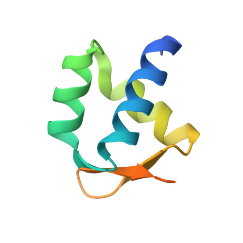
Since their initial characterization over 30 years ago, it has been believed that the archaeal B-family DNA polymerases are single-subunit enzymes. This contrasts with the multi-subunit B-family replicative polymerases of eukaryotes. Here we reveal that the highly studied PolB1 from Sulfolobus solfataricus exists as a heterotrimeric complex in cell extracts. Two small subunits, PBP1 and PBP2, associate with distinct surfaces of the larger catalytic subunit and influence the enzymatic properties of the DNA polymerase. Thus, multi-subunit replicative DNA polymerase holoenzymes are present in all three domains of life. We reveal the architecture of the assembly by a combination of cross-linking coupled with mass spectrometry, X-ray crystallography and single-particle electron microscopy. The small subunits stabilize the holoenzyme assembly and the acidic tail of one small subunit mitigates the ability of the enzyme to perform strand-displacement synthesis, with important implications for lagging strand DNA synthesis.
- Department of Molecular and Cellular Biochemistry, Indiana University, Simon Hall MSB, 212 S Hawthorne Dr, Bloomington, Indiana 47405, USA.
Organizational Affiliation: