5N2K | pdb_00005n2k

Structure of unbound Briakinumab FAb


Experimental Data Snapshot

  • Method: X-RAY DIFFRACTION
  • Resolution: 2.22 Å
  • R-Value Free: 
    0.234 (Depositor), 0.235 (DCC) 
  • R-Value Work: 
    0.204 (Depositor), 0.205 (DCC) 
  • R-Value Observed: 
    0.204 (Depositor) 

Starting Model: in silico
View more details

wwPDB Validation 3D Report Full Report

Validation slider image for 5N2K

This is version 2.2 of the entry. See complete history. 

Literature

Structural Activation of Pro-inflammatory Human Cytokine IL-23 by Cognate IL-23 Receptor Enables Recruitment of the Shared Receptor IL-12R beta 1.

Bloch, Y., Bouchareychas, L., Merceron, R., Skladanowska, K., Van den Bossche, L., Detry, S., Govindarajan, S., Elewaut, D., Haerynck, F., Dullaers, M., Adamopoulos, I.E., Savvides, S.N.

(2018) Immunity 48: 45-58.e6

  • DOI: https://doi.org/10.1016/j.immuni.2017.12.008
  • Primary Citation Related Structures: 
    5MXA, 5MZV, 5N2K, 5NJD

  • PubMed Abstract: 

    Interleukin-23 (IL-23), an IL-12 family cytokine, plays pivotal roles in pro-inflammatory T helper 17 cell responses linked to autoimmune and inflammatory diseases. Despite intense therapeutic targeting, structural and mechanistic insights into receptor complexes mediated by IL-23, and by IL-12 family members in general, have remained elusive. We determined a crystal structure of human IL-23 in complex with its cognate receptor, IL-23R, and revealed that IL-23R bound to IL-23 exclusively via its N-terminal immunoglobulin domain. The structural and functional hotspot of this interaction partially restructured the helical IL-23p19 subunit of IL-23 and restrained its IL-12p40 subunit to cooperatively bind the shared receptor IL-12Rβ1 with high affinity. Together with structural insights from the interaction of IL-23 with the inhibitory antibody briakinumab and by leveraging additional IL-23:antibody complexes, we propose a mechanistic paradigm for IL-23 and IL-12 whereby cognate receptor binding to the helical cytokine subunits primes recruitment of the shared receptors via the IL-12p40 subunit.


  • Organizational Affiliation: 
    • Laboratory for Protein Biochemistry and Biomolecular Engineering, Department of Biochemistry and Microbiology, Ghent University, 9052 Ghent, Belgium; VIB-UGent Center for Inflammation Research, 9052 Ghent, Belgium.

Macromolecule Content 

  • Total Structure Weight: 453.19 kDa 
  • Atom Count: 26,312 
  • Modeled Residue Count: 3,382 
  • Deposited Residue Count: 4,272 
  • Unique protein chains: 4

Macromolecules

Find similar proteins by:|  3D Structure
Entity ID: 1
MoleculeChains  Sequence LengthOrganismDetailsImage
Briakinumab FAb light chain
A,
C,
E,
I,
K,
M,
P [auth O]
245Homo sapiensMutation(s): 0 
Entity Groups
Sequence Clusters30% Identity50% Identity70% Identity90% Identity95% Identity100% Identity
Sequence Annotations
Expand
Reference Sequence
Find similar proteins by:|  3D Structure
Entity ID: 2
MoleculeChains  Sequence LengthOrganismDetailsImage
Briakinumab FAb heavy chain
B,
D,
F,
H,
L,
N,
O [auth P]
289Homo sapiensMutation(s): 0 
Entity Groups
Sequence Clusters30% Identity50% Identity70% Identity90% Identity95% Identity100% Identity
Sequence Annotations
Expand
Reference Sequence
Find similar proteins by:|  3D Structure
Entity ID: 3
MoleculeChains  Sequence LengthOrganismDetailsImage
Briakinumab FAb light chain245Homo sapiensMutation(s): 0 
Entity Groups
Sequence Clusters30% Identity50% Identity70% Identity90% Identity95% Identity100% Identity
Sequence Annotations
Expand
Reference Sequence
Find similar proteins by:|  3D Structure
Entity ID: 4
MoleculeChains  Sequence LengthOrganismDetailsImage
Briakinumab FAb heavy chain289Homo sapiensMutation(s): 0 
Entity Groups
Sequence Clusters30% Identity50% Identity70% Identity90% Identity95% Identity100% Identity
Sequence Annotations
Expand
Reference Sequence

Small Molecules

Ligands 1 Unique
IDChains Name / Formula / InChI Key2D Diagram3D Interactions
EDO

Query on EDO


Download:Ideal Coordinates CCD File
Q [auth I]1,2-ETHANEDIOL
C2 H6 O2
LYCAIKOWRPUZTN-UHFFFAOYSA-N
Modified Residues  1 Unique
IDChains Modified ResidueModification TypeFormula2D Diagram
PCA
Query on PCA
G, J
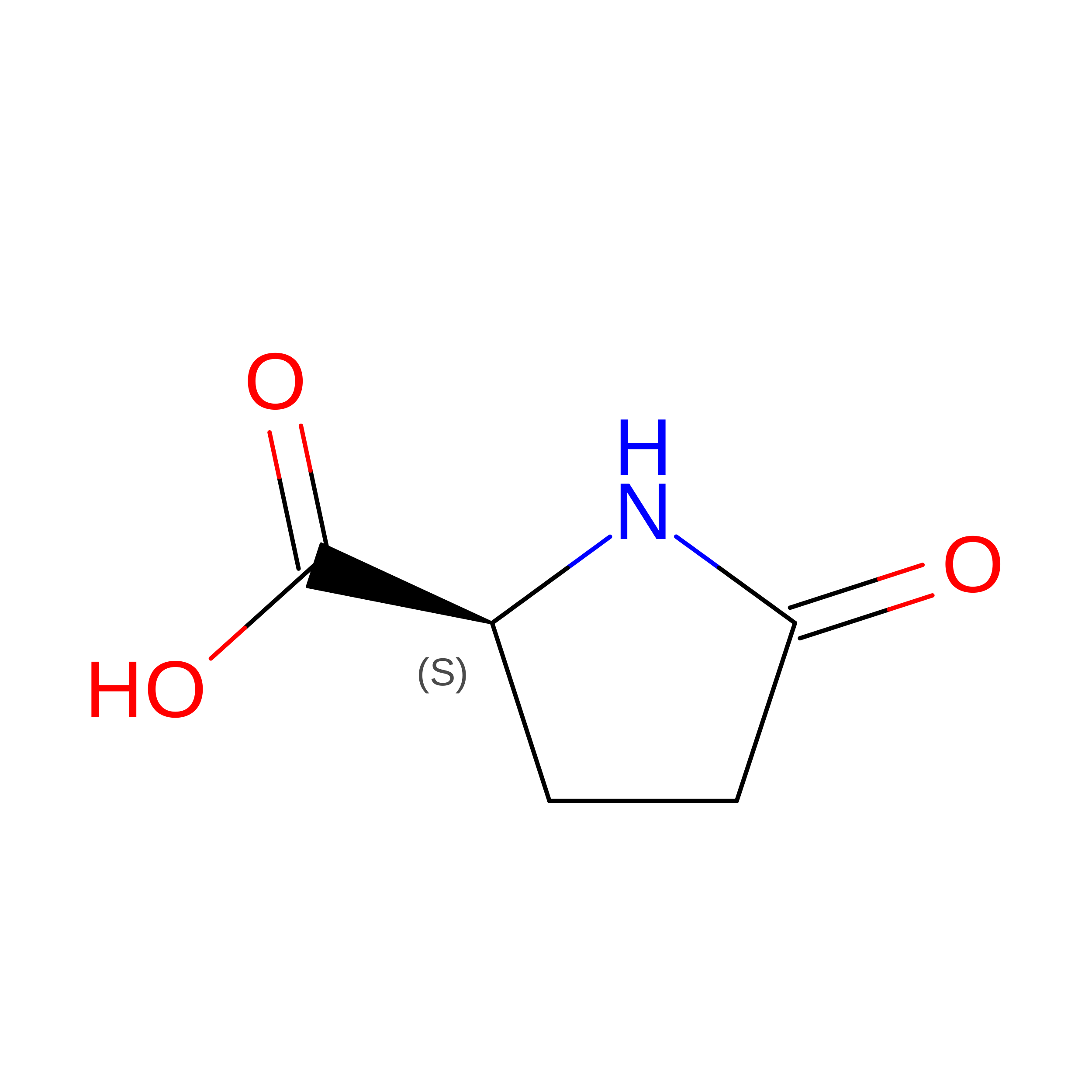
GLNPyrrolidone carboxylic acidC5 H7 N O3

Experimental Data & Validation

Experimental Data

  • Method: X-RAY DIFFRACTION
  • Resolution: 2.22 Å
  • R-Value Free:  0.234 (Depositor), 0.235 (DCC) 
  • R-Value Work:  0.204 (Depositor), 0.205 (DCC) 
  • R-Value Observed: 0.204 (Depositor) 
Space Group: P 1 21 1
Unit Cell:
Length ( Å )Angle ( ˚ )
a = 85.25α = 90
b = 172.54β = 106.16
c = 138.159γ = 90
Software Package:
Software NamePurpose
PHENIXrefinement
XDSdata reduction
XDSdata scaling
PHASERphasing

Structure Validation

View Full Validation Report


Entry History 

& Funding Information

Deposition Data


Funding OrganizationLocationGrant Number
VLAIOBelgium--

Revision History  (Full details and data files)

  • Version 1.0: 2018-01-03
    Type: Initial release
  • Version 1.1: 2018-01-10
    Changes: Database references
  • Version 1.2: 2018-01-31
    Changes: Database references
  • Version 2.0: 2020-03-11
    Changes: Polymer sequence
  • Version 2.1: 2024-05-01
    Changes: Data collection, Database references, Refinement description
  • Version 2.2: 2024-11-06
    Changes: Structure summary