5N2I | pdb_00005n2i

F420:NADPH oxidoreductase from Thermobifida fusca with NADP+ bound


Experimental Data Snapshot

  • Method: X-RAY DIFFRACTION
  • Resolution: 1.80 Å
  • R-Value Free: 
    0.214 (Depositor), 0.210 (DCC) 
  • R-Value Work: 
    0.165 (Depositor) 
  • R-Value Observed: 
    0.167 (Depositor) 

Starting Model: experimental
View more details

wwPDB Validation 3D Report Full Report

Validation slider image for 5N2I

Ligand Structure Quality Assessment 


This is version 1.2 of the entry. See complete history

Literature

Isolation and characterization of a thermostable F420:NADPH oxidoreductase from Thermobifida fusca.

Kumar, H.Nguyen, Q.T.Binda, C.Mattevi, A.Fraaije, M.W.

(2017) J Biological Chem 292: 10123-10130

  • DOI: https://doi.org/10.1074/jbc.M117.787754
  • Primary Citation Related Structures: 
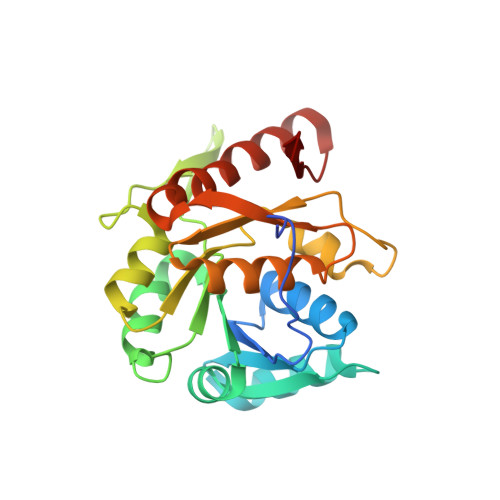
    5N2I

  • PubMed Abstract: 

    F 420 H 2 -dependent enzymes reduce a wide range of substrates that are otherwise recalcitrant to enzyme-catalyzed reduction, and their potential for applications in biocatalysis has attracted increasing attention. Thermobifida fusca is a moderately thermophilic bacterium and holds high biocatalytic potential as a source for several highly thermostable enzymes. We report here on the isolation and characterization of a thermostable F 420 : NADPH oxidoreductase (Tfu-FNO) from T. fusca , the first F 420 -dependent enzyme described from this bacterium. Tfu-FNO was heterologously expressed in Escherichia coli , yielding up to 200 mg of recombinant enzyme per liter of culture. We found that Tfu-FNO is highly thermostable, reaching its highest activity at 65 °C and that Tfu-FNO is likely to act in vivo as an F 420 reductase at the expense of NADPH, similar to its counterpart in Streptomyces griseus We obtained the crystal structure of FNO in complex with NADP + at 1.8 Å resolution, providing the first bacterial FNO structure. The overall architecture and NADP + -binding site of Tfu-FNO were highly similar to those of the Archaeoglobus fulgidus FNO (Af-FNO). The active site is located in a hydrophobic pocket between an N-terminal dinucleotide binding domain and a smaller C-terminal domain. Residues interacting with the 2'-phosphate of NADP + were probed by targeted mutagenesis, indicating that Thr-28, Ser-50, Arg-51, and Arg-55 are important for discriminating between NADP + and NAD + Interestingly, a T28A mutant increased the kinetic efficiency >3-fold as compared with the wild-type enzyme when NADH is the substrate. The biochemical and structural data presented here provide crucial insights into the molecular recognition of the two cofactors, F 420 and NAD(P)H by FNO.


  • Organizational Affiliation
    • From the Molecular Enzymology Group, Groningen Biomolecular Sciences and Biotechnology Institute, University of Groningen, Nijenborgh 4, 9747 AG Groningen, The Netherlands.

Macromolecule Content 

  • Total Structure Weight: 100.95 kDa 
  • Atom Count: 7,428 
  • Modeled Residue Count: 893 
  • Deposited Residue Count: 928 
  • Unique protein chains: 1

Macromolecules

Find similar proteins by:|  3D Structure
Entity ID: 1
MoleculeChains  Sequence LengthOrganismDetailsImage
Reduced coenzyme F420:NADP oxidoreductase
A, B, C, D
232Thermobifida fusca YXMutation(s): 0 
Gene Names: Tfu_0970
EC: 1.6.99
UniProt
Find proteins for Q47RA9 (Thermobifida fusca (strain YX))
Explore Q47RA9 
Go to UniProtKB:  Q47RA9
Entity Groups
Sequence Clusters30% Identity50% Identity70% Identity90% Identity95% Identity100% Identity
UniProt GroupQ47RA9
Sequence Annotations
Expand
Reference Sequence

Small Molecules

Ligands 2 Unique
IDChains Name / Formula / InChI Key2D Diagram3D Interactions
NAP

Query on NAP


Download:Ideal Coordinates CCD File
E [auth A],
J [auth B],
L [auth C],
N [auth D]
NADP NICOTINAMIDE-ADENINE-DINUCLEOTIDE PHOSPHATE
C21 H28 N7 O17 P3
XJLXINKUBYWONI-NNYOXOHSSA-N
GOL

Query on GOL


Download:Ideal Coordinates CCD File
F [auth A]
G [auth A]
H [auth A]
I [auth A]
K [auth B]
F [auth A],
G [auth A],
H [auth A],
I [auth A],
K [auth B],
M [auth C],
O [auth D]
GLYCEROL
C3 H8 O3
PEDCQBHIVMGVHV-UHFFFAOYSA-N

Experimental Data & Validation

Experimental Data

  • Method: X-RAY DIFFRACTION
  • Resolution: 1.80 Å
  • R-Value Free:  0.214 (Depositor), 0.210 (DCC) 
  • R-Value Work:  0.165 (Depositor) 
  • R-Value Observed: 0.167 (Depositor) 
Space Group: P 21 21 21
Unit Cell:
Length ( Å )Angle ( ˚ )
a = 82.389α = 90
b = 86.089β = 90
c = 136.775γ = 90
Software Package:
Software NamePurpose
REFMACrefinement
XDSdata reduction
Aimlessdata scaling
PHASERphasing

Structure Validation

View Full Validation Report


Ligand Structure Quality Assessment 


Entry History 

& Funding Information

Deposition Data


Funding OrganizationLocationGrant Number
European CommissionNetherlandsErasmus Mundus Action 2 project for Lot11 - Svaagata.eu
University of GroningenNetherlandsUbbo Emius scholarship

Revision History  (Full details and data files)

  • Version 1.0: 2017-04-26
    Type: Initial release
  • Version 1.1: 2017-06-28
    Changes: Data collection, Database references
  • Version 1.2: 2024-01-17
    Changes: Data collection, Database references, Refinement description