5N26

X-ray structure of human heavy chain ferritin in complex with cisplatin


Experimental Data Snapshot

  • Method: X-RAY DIFFRACTION
  • Resolution: 2.05 Å
  • R-Value Free: 0.206 
  • R-Value Work: 0.163 
  • R-Value Observed: 0.165 

wwPDB Validation   3D Report Full Report


This is version 1.1 of the entry. See complete history


Literature

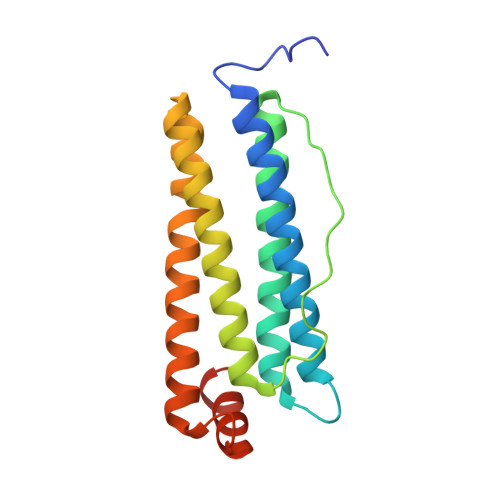
Cisplatin Binding Sites in Human H-Chain Ferritin.

Ferraro, G.Ciambellotti, S.Messori, L.Merlino, A.

(2017) Inorg Chem 56: 9064-9070

  • DOI: https://doi.org/10.1021/acs.inorgchem.7b01072
  • Primary Citation of Related Structures:  
    5N26, 5N27

  • PubMed Abstract: 
  • The aim of this work is to identify the cisplatin binding sites on human H-chain ferritin. High-resolution X-ray crystallography reveals that cisplatin binds four distinct protein sites, that is, the side chains of His136 and Lys68, the side chain of His105, the side chain of Cys90 and the side chain of Cys102 ...

    The aim of this work is to identify the cisplatin binding sites on human H-chain ferritin. High-resolution X-ray crystallography reveals that cisplatin binds four distinct protein sites, that is, the side chains of His136 and Lys68, the side chain of His105, the side chain of Cys90 and the side chain of Cys102. These Pt binding sites are compared with those observed for the adduct that cisplatin forms upon encapsulation within horse spleen L-chain ferritin (87% identity with human L-chain ferritin).


    Organizational Affiliation

    Institute of Biostructures and Bioimages , Naples, Italy.


Macromolecules
Find similar proteins by:  (by identity cutoff)  |  3D Structure
Entity ID: 1
MoleculeChainsSequence LengthOrganismDetailsImage
Ferritin heavy chain182Homo sapiensMutation(s): 0 
Gene Names: FTH1FTHFTHL6OK/SW-cl.84PIG15
EC: 1.16.3.1
UniProt & NIH Common Fund Data Resources
Find proteins for P02794 (Homo sapiens)
Explore P02794 
Go to UniProtKB:  P02794
PHAROS:  P02794
Entity Groups  
Sequence Clusters30% Identity50% Identity70% Identity90% Identity95% Identity100% Identity
UniProt GroupP02794
Protein Feature View
Expand
  • Reference Sequence
Small Molecules
Ligands 4 Unique
IDChainsName / Formula / InChI Key2D Diagram3D Interactions
CPT
Query on CPT

Download Ideal Coordinates CCD File 
B [auth A],
D [auth A],
E [auth A]
Cisplatin
Cl2 H6 N2 Pt
LXZZYRPGZAFOLE-UHFFFAOYSA-L
 Ligand Interaction
73M
Query on 73M

Download Ideal Coordinates CCD File 
C [auth A]bis(azanyl)-chloranyl-oxidanyl-platinum
Cl H5 N2 O Pt
DOKMFIMZADYDDQ-UHFFFAOYSA-L
 Ligand Interaction
CL
Query on CL

Download Ideal Coordinates CCD File 
F [auth A]
G [auth A]
H [auth A]
I [auth A]
J [auth A]
F [auth A],
G [auth A],
H [auth A],
I [auth A],
J [auth A],
K [auth A]
CHLORIDE ION
Cl
VEXZGXHMUGYJMC-UHFFFAOYSA-M
 Ligand Interaction
MG
Query on MG

Download Ideal Coordinates CCD File 
L [auth A],
M [auth A],
N [auth A],
O [auth A],
P [auth A]
MAGNESIUM ION
Mg
JLVVSXFLKOJNIY-UHFFFAOYSA-N
 Ligand Interaction
Experimental Data & Validation

Experimental Data

  • Method: X-RAY DIFFRACTION
  • Resolution: 2.05 Å
  • R-Value Free: 0.206 
  • R-Value Work: 0.163 
  • R-Value Observed: 0.165 
  • Space Group: F 4 3 2
Unit Cell:
Length ( Å )Angle ( ˚ )
a = 183.069α = 90
b = 183.069β = 90
c = 183.069γ = 90
Software Package:
Software NamePurpose
REFMACrefinement
HKL-2000data reduction
HKL-2000data scaling
PHASERphasing

Structure Validation

View Full Validation Report


Entry History 

Deposition Data

Revision History  (Full details and data files)

  • Version 1.0: 2017-08-02
    Type: Initial release
  • Version 1.1: 2017-08-16
    Changes: Database references