Convergent evolution involving dimeric and trimeric dUTPases in pathogenicity island mobilization.
Donderis, J., Bowring, J., Maiques, E., Ciges-Tomas, J.R., Alite, C., Mehmedov, I., Tormo-Mas, M.A., Penades, J.R., Marina, A.(2017) PLoS Pathog 13: e1006581-e1006581
- PubMed: 28892519 Search on PubMedSearch on PubMed Central
- DOI: https://doi.org/10.1371/journal.ppat.1006581
- Primary Citation Related Structures:
5MYD, 5MYF, 5MYI - PubMed Abstract:
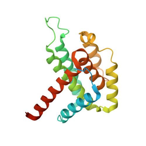
The dUTPase (Dut) enzymes, encoded by almost all free-living organisms and some viruses, prevent the misincorporation of uracil into DNA. We previously proposed that trimeric Duts are regulatory proteins involved in different cellular processes; including the phage-mediated transfer of the Staphylococcus aureus pathogenicity island SaPIbov1. Recently, it has been shown that the structurally unrelated dimeric Dut encoded by phage ϕNM1 is similarly able to mobilize SaPIbov1, suggesting dimeric Duts could also be regulatory proteins. How this is accomplished remains unsolved. Here, using in vivo, biochemical and structural approaches, we provide insights into the signaling mechanism used by the dimeric Duts to induce the SaPIbov1 cycle. As reported for the trimeric Duts, dimeric Duts contain an extremely variable region, here named domain VI, which is involved in the regulatory capacity of these enzymes. Remarkably, our results also show that the dimeric Dut signaling mechanism is modulated by dUTP, as with the trimeric Duts. Overall, our results demonstrate that although unrelated both in sequence and structure, dimeric and trimeric Duts control SaPI transfer by analogous mechanisms, representing a fascinating example of convergent evolution. This conserved mode of action highlights the biological significance of Duts as regulatory molecules.
- Instituto de Biomedicina de Valencia (IBV-CSIC) and CIBER de Enfermedades Raras (CIBERER), Valencia, Spain.
Organizational Affiliation: