5MTZ | pdb_00005mtz

Crystal structure of a long form RNase Z from yeast


Experimental Data Snapshot

  • Method: X-RAY DIFFRACTION
  • Resolution: 2.99 Å
  • R-Value Free: 
    0.262 (Depositor), 0.243 (DCC) 
  • R-Value Work: 
    0.181 (Depositor) 
  • R-Value Observed: 
    0.185 (Depositor) 

wwPDB Validation 3D Report Full Report

Validation slider image for 5MTZ

This is version 1.3 of the entry. See complete history

Literature

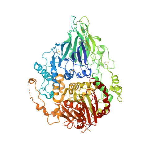
The crystal structure of Trz1, the long form RNase Z from yeast.

Ma, M.Li de la Sierra-Gallay, I.Lazar, N.Pellegrini, O.Durand, D.Marchfelder, A.Condon, C.van Tilbeurgh, H.

(2017) Nucleic Acids Res 45: 6209-6216

  • DOI: https://doi.org/10.1093/nar/gkx216
  • Primary Citation Related Structures: 
    5MTZ

  • PubMed Abstract: 

    tRNAs are synthesized as precursor RNAs that have to undergo processing steps to become functional. Yeast Trz1 is a key endoribonuclease involved in the 3΄ maturation of tRNAs in all domains of life. It is a member of the β-lactamase family of RNases, characterized by an HxHxDH sequence motif involved in coordination of catalytic Zn-ions. The RNase Z family consists of two subfamilies: the short (250-400 residues) and the long forms (about double in size). Short form RNase Z enzymes act as homodimers: one subunit embraces tRNA with a protruding arm, while the other provides the catalytic site. The long form is thought to contain two fused β-lactamase domains within a single polypeptide. Only structures of short form RNase Z enzymes are known. Here we present the 3.1 Å crystal structure of the long-form Trz1 from Saccharomyces cerevisiae. Trz1 is organized into two β-lactamase domains connected by a long linker. The N-terminal domain has lost its catalytic residues, but retains the long flexible arm that is important for tRNA binding, while it is the other way around in the C-terminal domain. Trz1 likely evolved from a duplication and fusion of the gene encoding the monomeric short form RNase Z.


  • Organizational Affiliation
    • Institute for Integrative Biology of the Cell (I2BC), CEA, CNRS UMR 9198, University of Paris-Sud, Université Paris-Saclay, 91198 Gif sur Yvette Cedex, France.

Macromolecule Content 

  • Total Structure Weight: 204.48 kDa 
  • Atom Count: 12,472 
  • Modeled Residue Count: 1,523 
  • Deposited Residue Count: 1,748 
  • Unique protein chains: 1

Macromolecules

Find similar proteins by:|  3D Structure
Entity ID: 1
MoleculeChains  Sequence LengthOrganismDetailsImage
Ribonuclease Z
A, B
874Saccharomyces cerevisiaeMutation(s): 0 
Gene Names: TRZ1YKR079C
EC: 3.1.26.11
UniProt
Find proteins for P36159 (Saccharomyces cerevisiae (strain ATCC 204508 / S288c))
Explore P36159 
Go to UniProtKB:  P36159
Entity Groups
Sequence Clusters30% Identity50% Identity70% Identity90% Identity95% Identity100% Identity
UniProt GroupP36159
Sequence Annotations
Expand
Reference Sequence

Experimental Data & Validation

Experimental Data

  • Method: X-RAY DIFFRACTION
  • Resolution: 2.99 Å
  • R-Value Free:  0.262 (Depositor), 0.243 (DCC) 
  • R-Value Work:  0.181 (Depositor) 
  • R-Value Observed: 0.185 (Depositor) 
Space Group: P 41
Unit Cell:
Length ( Å )Angle ( ˚ )
a = 136α = 90
b = 136β = 90
c = 115.81γ = 90
Software Package:
Software NamePurpose
REFMACrefinement
XSCALEdata scaling
PDB_EXTRACTdata extraction
XDSdata scaling
XDSdata reduction
SHELXCDphasing
PHASERphasing
BUCCANEERmodel building

Structure Validation

View Full Validation Report


Entry History 

& Funding Information

Deposition Data


Funding OrganizationLocationGrant Number
China Scholarship CouncilChina201206180025
French Infrastructure for Integrates Structural Biology (FRISBI)FranceANR-10-INSB-05

Revision History  (Full details and data files)

  • Version 1.0: 2017-06-21
    Type: Initial release
  • Version 1.1: 2018-01-17
    Changes: Structure summary
  • Version 1.2: 2018-01-31
    Changes: Database references
  • Version 1.3: 2024-11-06
    Changes: Data collection, Database references, Derived calculations, Structure summary