5MS4 | pdb_00005ms4

Kallikrein-related peptidase 8 leupeptin inhibitor complex


Experimental Data Snapshot

  • Method: X-RAY DIFFRACTION
  • Resolution: 2.10 Å
  • R-Value Free: 
    0.259 (Depositor), 0.251 (DCC) 
  • R-Value Work: 
    0.212 (Depositor), 0.215 (DCC) 
  • R-Value Observed: 
    0.239 (Depositor) 

wwPDB Validation 3D Report Full Report

Validation slider image for 5MS4

This is version 2.2 of the entry. See complete history

Literature

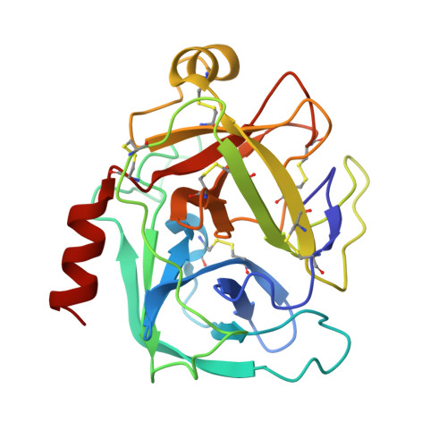
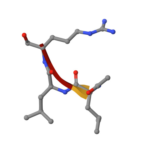
Structural determinants of specificity and regulation of activity in the allosteric loop network of human KLK8/neuropsin.

Debela, M.Magdolen, V.Skala, W.Elsasser, B.Schneider, E.L.Craik, C.S.Biniossek, M.L.Schilling, O.Bode, W.Brandstetter, H.Goettig, P.

(2018) Sci Rep 8: 10705-10705

  • DOI: https://doi.org/10.1038/s41598-018-29058-6
  • Primary Citation Related Structures: 
    5MS4

  • PubMed Abstract: 

    Human KLK8/neuropsin, a kallikrein-related serine peptidase, is mostly expressed in skin and the hippocampus regions of the brain, where it regulates memory formation by synaptic remodeling. Substrate profiles of recombinant KLK8 were analyzed with positional scanning using fluorogenic tetrapeptides and the proteomic PICS approach, which revealed the prime side specificity. Enzyme kinetics with optimized substrates showed stimulation by Ca 2+ and inhibition by Zn 2+ , which are physiological regulators. Crystal structures of KLK8 with a ligand-free active site and with the inhibitor leupeptin explain the subsite specificity and display Ca 2+ bound to the 75-loop. The variants D70K and H99A confirmed the antagonistic role of the cation binding sites. Molecular docking and dynamics calculations provided insights in substrate binding and the dual regulation of activity by Ca 2+ and Zn 2+ , which are important in neuron and skin physiology. Both cations participate in the allosteric surface loop network present in related serine proteases. A comparison of the positional scanning data with substrates from brain suggests an adaptive recognition by KLK8, based on the tertiary structures of its targets. These combined findings provide a comprehensive picture of the molecular mechanisms underlying the enzyme activity of KLK8.


  • Organizational Affiliation
    • Max-Planck-Institut für Biochemie, Proteinase Research Group, 82152, Martinsried, Germany.

Macromolecule Content 

  • Total Structure Weight: 100.04 kDa 
  • Atom Count: 8,120 
  • Modeled Residue Count: 928 
  • Deposited Residue Count: 928 
  • Unique protein chains: 2

Macromolecules

Find similar proteins by:|  3D Structure
Entity ID: 1
MoleculeChains  Sequence LengthOrganismDetailsImage
Kallikrein-8
A, B, C, D
228Homo sapiensMutation(s): 0 
Gene Names: KLK8NRPNPRSS19TADG14UNQ283/PRO322
EC: 3.4.21.118
UniProt & NIH Common Fund Data Resources
Find proteins for O60259 (Homo sapiens)
Explore O60259 
Go to UniProtKB:  O60259
PHAROS:  O60259
GTEx:  ENSG00000129455 
Entity Groups
Sequence Clusters30% Identity50% Identity70% Identity90% Identity95% Identity100% Identity
UniProt GroupO60259
Sequence Annotations
Expand
Reference Sequence
Find similar proteins by:  Sequence   |   3D Structure  
Entity ID: 2
MoleculeChains  Sequence LengthOrganismDetailsImage
LEUPEPTIN
E, F, G, H
4synthetic constructMutation(s): 0 

Small Molecules

Modified Residues  1 Unique
IDChains TypeFormula2D DiagramParent
AR7
Query on AR7
E, F, G, H
PEPTIDE-LIKEC6 H17 N4 O2ARG

Experimental Data & Validation

Experimental Data

  • Method: X-RAY DIFFRACTION
  • Resolution: 2.10 Å
  • R-Value Free:  0.259 (Depositor), 0.251 (DCC) 
  • R-Value Work:  0.212 (Depositor), 0.215 (DCC) 
  • R-Value Observed: 0.239 (Depositor) 
Space Group: P 1 21 1
Unit Cell:
Length ( Å )Angle ( ˚ )
a = 83.103α = 90
b = 46.045β = 91.4
c = 103.349γ = 90
Software Package:
Software NamePurpose
PHENIXrefinement
XDSdata reduction
SCALAdata scaling
PHASERphasing
PDB_EXTRACTdata extraction

Structure Validation

View Full Validation Report


Entry History 

& Funding Information

Deposition Data


Funding OrganizationLocationGrant Number
Austrian Science FundAustriaP25003-B21

Revision History  (Full details and data files)

  • Version 1.0: 2018-01-17
    Type: Initial release
  • Version 1.1: 2018-08-01
    Changes: Data collection, Database references
  • Version 2.0: 2019-09-18
    Changes: Advisory, Atomic model, Data collection, Database references, Derived calculations, Non-polymer description, Polymer sequence, Source and taxonomy, Structure summary
  • Version 2.1: 2019-10-09
    Changes: Data collection, Derived calculations
  • Version 2.2: 2024-10-09
    Changes: Data collection, Database references, Derived calculations, Structure summary