Mechanistic insights into the allosteric regulation of bacterial ADP-glucose pyrophosphorylases.
Comino, N., Cifuente, J.O., Marina, A., Orrantia, A., Eguskiza, A., Guerin, M.E.(2017) J Biological Chem 292: 6255-6268
- PubMed: 28223362 Search on PubMedSearch on PubMed Central
- DOI: https://doi.org/10.1074/jbc.M116.773408
- Primary Citation Related Structures:
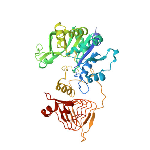
5MNI - PubMed Abstract:
ADP-glucose pyrophosphorylase (AGPase) controls bacterial glycogen and plant starch biosynthetic pathways, the most common carbon storage polysaccharides in nature. AGPase activity is allosterically regulated by a series of metabolites in the energetic flux within the cell. Very recently, we reported the first crystal structures of the paradigmatic AGPase from Escherichia coli ( Ec AGPase) in complex with its preferred physiological negative and positive allosteric regulators, adenosine 5'-monophosphate (AMP) and fructose 1,6-bisphosphate (FBP), respectively. However, understanding the molecular mechanism by which AMP and FBP allosterically modulates Ec AGPase enzymatic activity still remains enigmatic. Here we found that single point mutations of key residues in the AMP-binding site decrease its inhibitory effect but also clearly abolish the overall AMP-mediated stabilization effect in wild-type Ec AGPase. Single point mutations of key residues for FBP binding did not revert the AMP-mediated stabilization. Strikingly, an Ec AGPase-R130A mutant displayed a dramatic increase in activity when compared with wild-type Ec AGPase, and this increase correlated with a significant increment of glycogen content in vivo The crystal structure of Ec AGPase-R130A revealed unprecedented conformational changes in structural elements involved in the allosteric signal transmission. Altogether, we propose a model in which the positive and negative energy reporters regulate AGPase catalytic activity via intra- and interprotomer cross-talk, with a "sensory motif" and two loops, RL1 and RL2, flanking the ATP-binding site playing a significant role. The information reported herein provides exciting possibilities for industrial/biotechnological applications.
- From the Structural Biology Unit, CIC bioGUNE, Bizkaia Technology Park, 48160 Derio, Spain.
Organizational Affiliation: