5MG5 | pdb_00005mg5

A multi-component acyltransferase PhlABC from Pseudomonas protegens soaked with the monoacetylphloroglucinol (MAPG)


Experimental Data Snapshot

  • Method: X-RAY DIFFRACTION
  • Resolution: 3.44 Å
  • R-Value Free: 
    0.221 (Depositor), 0.222 (DCC) 
  • R-Value Work: 
    0.165 (Depositor), 0.168 (DCC) 
  • R-Value Observed: 
    0.168 (Depositor) 

Starting Model: experimental
View more details

wwPDB Validation 3D Report Full Report

Validation slider image for 5MG5

This is version 1.4 of the entry. See complete history

Literature

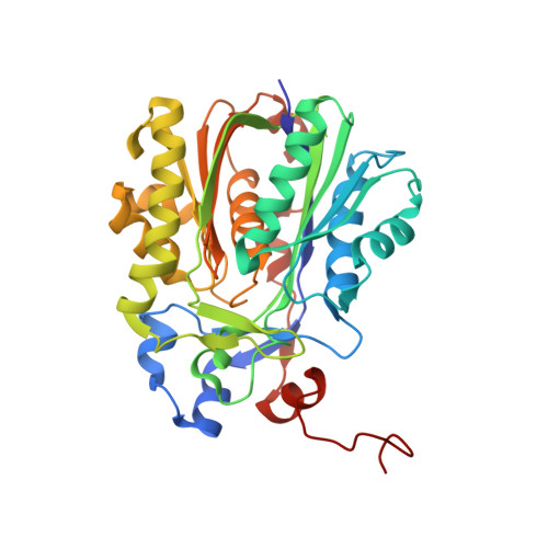
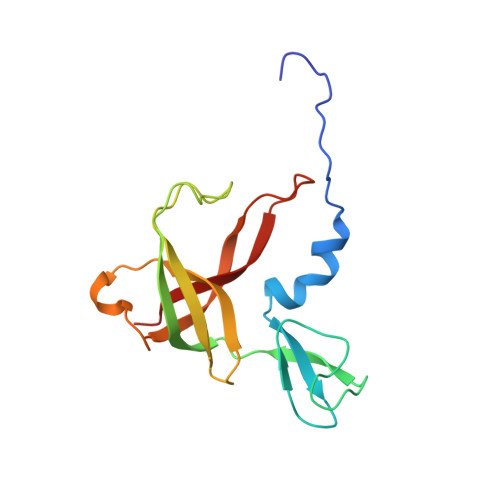
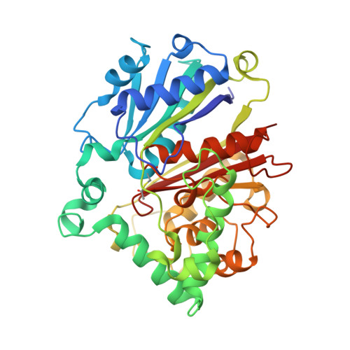
Structure and Catalytic Mechanism of a Bacterial Friedel-Crafts Acylase.

Pavkov-Keller, T.Schmidt, N.G.Zadlo-Dobrowolska, A.Kroutil, W.Gruber, K.

(2019) Chembiochem 20: 88-95

  • DOI: https://doi.org/10.1002/cbic.201800462
  • Primary Citation Related Structures: 
    5M3K, 5MG5

  • PubMed Abstract: 

    C-C bond-forming reactions are key transformations for setting up the carbon frameworks of organic compounds. In this context, Friedel-Crafts acylation is commonly used for the synthesis of aryl ketones, which are common motifs in many fine chemicals and natural products. A bacterial multicomponent acyltransferase from Pseudomonas protegens (PpATase) catalyzes such Friedel-Crafts C-acylation of phenolic substrates in aqueous solution, reaching up to >99 % conversion without the need for CoA-activated reagents. We determined X-ray crystal structures of the native and ligand-bound complexes. This multimeric enzyme consists of three subunits: PhlA, PhlB, and PhlC, arranged in a Phl(A 2 C 2 ) 2 B 4 composition. The structure of a reaction intermediate obtained from crystals soaked with the natural substrate 1-(2,4,6-trihydroxyphenyl)ethanone together with site-directed mutagenesis studies revealed that only residues from the PhlC subunits are involved in the acyl transfer reaction, with Cys88 very likely playing a significant role during catalysis. These structural and mechanistic insights form the basis of further enzyme engineering efforts directed towards enhancing the substrate scope of this enzyme.


  • Organizational Affiliation
    • Austrian Centre of Industrial Biotechnology (ACIB), Petersgasse 14, 8010, Graz, Austria.

Macromolecule Content 

  • Total Structure Weight: 783.54 kDa 
  • Atom Count: 54,522 
  • Modeled Residue Count: 7,194 
  • Deposited Residue Count: 7,248 
  • Unique protein chains: 3

Macromolecules

Find similar proteins by:|  3D Structure
Entity ID: 1
MoleculeChains  Sequence LengthOrganismDetailsImage
Hydroxymethylglutaryl-CoA synthase
A, D, G, J, M
A, D, G, J, M, P, S, V
362Pseudomonas protegensMutation(s): 0 
Gene Names: CEP86_09500
Find similar proteins by:|  3D Structure
Entity ID: 2
MoleculeChains  Sequence LengthOrganismDetailsImage
2,4-diacetylphloroglucinol biosynthesis protein
B, E, H, K, N
B, E, H, K, N, Q, T, W
146Pseudomonas protegensMutation(s): 0 
Gene Names: CEP86_09490
Find similar proteins by:|  3D Structure
Entity ID: 3
MoleculeChains  Sequence LengthOrganismDetailsImage
2,4-diacetylphloroglucinol biosynthesis protein PhlC
C, F, I, L, O
C, F, I, L, O, R, U, X
398Pseudomonas protegens Pf-5Mutation(s): 0 
Gene Names: phlCPFL_5955
UniProt
Find proteins for Q4K420 (Pseudomonas fluorescens (strain ATCC BAA-477 / NRRL B-23932 / Pf-5))
Explore Q4K420 
Go to UniProtKB:  Q4K420
Entity Groups
Sequence Clusters30% Identity50% Identity70% Identity90% Identity95% Identity100% Identity
UniProt GroupQ4K420
Sequence Annotations
Expand
Reference Sequence

Small Molecules

Ligands 2 Unique
IDChains Name / Formula / InChI Key2D Diagram3D Interactions
13X

Query on 13X


Download:Ideal Coordinates CCD File
DA [auth L],
FA [auth O],
HA [auth R],
Z [auth C]
benzene-1,3,5-triol
C6 H6 O3
QCDYQQDYXPDABM-UHFFFAOYSA-N
ZN

Query on ZN


Download:Ideal Coordinates CCD File
AA [auth E]
BA [auth H]
CA [auth K]
EA [auth N]
GA [auth Q]
AA [auth E],
BA [auth H],
CA [auth K],
EA [auth N],
GA [auth Q],
IA [auth T],
JA [auth W],
Y [auth B]
ZINC ION
Zn
PTFCDOFLOPIGGS-UHFFFAOYSA-N
Modified Residues  1 Unique
IDChains TypeFormula2D DiagramParent
SCY
Query on SCY
C, F, I, L, O
C, F, I, L, O, R, U, X
L-PEPTIDE LINKINGC5 H9 N O3 SCYS

Experimental Data & Validation

Experimental Data

  • Method: X-RAY DIFFRACTION
  • Resolution: 3.44 Å
  • R-Value Free:  0.221 (Depositor), 0.222 (DCC) 
  • R-Value Work:  0.165 (Depositor), 0.168 (DCC) 
  • R-Value Observed: 0.168 (Depositor) 
Space Group: P 21 21 21
Unit Cell:
Length ( Å )Angle ( ˚ )
a = 104.793α = 90
b = 229.775β = 90
c = 311.129γ = 90
Software Package:
Software NamePurpose
PHENIXrefinement
XDSdata reduction
XDSdata scaling
PHASERphasing

Structure Validation

View Full Validation Report


Entry History 

Deposition Data

Revision History  (Full details and data files)

  • Version 1.0: 2017-12-20
    Type: Initial release
  • Version 1.1: 2018-10-31
    Changes: Data collection, Database references
  • Version 1.2: 2019-02-27
    Changes: Data collection, Database references
  • Version 1.3: 2024-01-17
    Changes: Data collection, Database references, Derived calculations, Refinement description
  • Version 1.4: 2024-10-09
    Changes: Structure summary