5MFT

The crystal structure of E. coli Aminopeptidase N in complex with 7-amino-1-bromo-4-phenyl-5,7,8,9-tetrahydrobenzocyclohepten-6-one


Experimental Data Snapshot

  • Method: X-RAY DIFFRACTION
  • Resolution: 1.59 Å
  • R-Value Free: 0.139 
  • R-Value Work: 0.111 
  • R-Value Observed: 0.111 

wwPDB Validation   3D Report Full Report


This is version 2.0 of the entry. See complete history


Literature

Insight into the remarkable affinity and selectivity of the aminobenzosuberone scaffold for the M1 aminopeptidases family based on structure analysis.

Peng, G.McEwen, A.G.Olieric, V.Schmitt, C.Albrecht, S.Cavarelli, J.Tarnus, C.

(2017) Proteins 85: 1413-1421

  • DOI: https://doi.org/10.1002/prot.25301
  • Primary Citation of Related Structures:  
    5MFR, 5MFS, 5MFT

  • PubMed Abstract: 

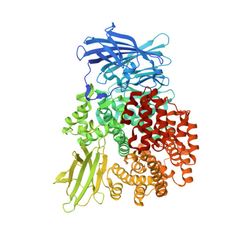
    Aminopeptidases are ubiquitous hydrolases that cleave the N-terminal residues of proteins and oligopeptides. They are broadly distributed throughout all kingdoms of life and have been implicated in a wide variety of physiological processes, including viral infection, parasite metabolism, protein processing, regulation of peptide hormones, and cancer cell proliferation. Members of the M1 family, also termed gluzincins, are defined by two highly conserved motifs in the catalytic domain: a zinc-binding motif, HEXXH-(X18)-E; and an exopeptidase motif, GXMEN. We report the high-resolution X-ray structures of E. coli aminopeptidase N (PepN) in complex with three aminobenzosuberone scaffolds that display various Ki values (50, 0.33, and 0.034 µM) and provide a compelling view of the outstanding selectivity of these chemical entities for the M1 aminopeptidases. This series of inhibitors interacts as transition state mimics with highly conserved residues of the catalytic machinery and substrate recognition sites. Structural comparisons and model-building studies allowed a deep interpretation of the SAR observed for bacterial, as well as mammalian enzymes. Proteins 2017; 85:1413-1421. © 2017 Wiley Periodicals, Inc.


  • Organizational Affiliation

    Paul Scherrer Institut (SLS) WSLB, 5232, Villigen, Suisse, Switzerland.


Macromolecules
Find similar proteins by:  (by identity cutoff)  |  3D Structure
Entity ID: 1
MoleculeChains Sequence LengthOrganismDetailsImage
Aminopeptidase N891Escherichia coli K-12Mutation(s): 0 
Gene Names: pepNb0932JW0915
EC: 3.4.11.2
UniProt
Find proteins for P04825 (Escherichia coli (strain K12))
Explore P04825 
Go to UniProtKB:  P04825
Entity Groups  
Sequence Clusters30% Identity50% Identity70% Identity90% Identity95% Identity100% Identity
UniProt GroupP04825
Sequence Annotations
Expand
  • Reference Sequence
Small Molecules
Ligands 7 Unique
IDChains Name / Formula / InChI Key2D Diagram3D Interactions
7MF
Query on 7MF

Download Ideal Coordinates CCD File 
C [auth A][(7~{S})-1-bromanyl-6,6-bis(oxidanyl)-4-phenyl-5,7,8,9-tetrahydrobenzo[7]annulen-7-yl]azanium
C17 H19 Br N O2
YMWOHZAVFJCVSC-INIZCTEOSA-O
MLI
Query on MLI

Download Ideal Coordinates CCD File 
V [auth A],
W [auth A],
X [auth A],
Y [auth A],
Z [auth A]
MALONATE ION
C3 H2 O4
OFOBLEOULBTSOW-UHFFFAOYSA-L
GOL
Query on GOL

Download Ideal Coordinates CCD File 
J [auth A]
K [auth A]
L [auth A]
M [auth A]
N [auth A]
J [auth A],
K [auth A],
L [auth A],
M [auth A],
N [auth A],
O [auth A],
P [auth A],
Q [auth A]
GLYCEROL
C3 H8 O3
PEDCQBHIVMGVHV-UHFFFAOYSA-N
DMS
Query on DMS

Download Ideal Coordinates CCD File 
R [auth A],
S [auth A]
DIMETHYL SULFOXIDE
C2 H6 O S
IAZDPXIOMUYVGZ-UHFFFAOYSA-N
ZN
Query on ZN

Download Ideal Coordinates CCD File 
B [auth A]ZINC ION
Zn
PTFCDOFLOPIGGS-UHFFFAOYSA-N
CL
Query on CL

Download Ideal Coordinates CCD File 
T [auth A],
U [auth A]
CHLORIDE ION
Cl
VEXZGXHMUGYJMC-UHFFFAOYSA-M
NA
Query on NA

Download Ideal Coordinates CCD File 
D [auth A]
E [auth A]
F [auth A]
G [auth A]
H [auth A]
D [auth A],
E [auth A],
F [auth A],
G [auth A],
H [auth A],
I [auth A]
SODIUM ION
Na
FKNQFGJONOIPTF-UHFFFAOYSA-N
Binding Affinity Annotations 
IDSourceBinding Affinity
7MF Binding MOAD:  5MFT Ki: 34 (nM) from 1 assay(s)
Experimental Data & Validation

Experimental Data

  • Method: X-RAY DIFFRACTION
  • Resolution: 1.59 Å
  • R-Value Free: 0.139 
  • R-Value Work: 0.111 
  • R-Value Observed: 0.111 
  • Space Group: P 31 2 1
Unit Cell:
Length ( Å )Angle ( ˚ )
a = 120.651α = 90
b = 120.651β = 90
c = 170.676γ = 120
Software Package:
Software NamePurpose
Aimlessdata scaling
PHENIXrefinement
PDB_EXTRACTdata extraction
XDSdata reduction
PHASERphasing
XSCALEdata scaling

Structure Validation

View Full Validation Report


Entry History & Funding Information

Deposition Data


Funding OrganizationLocationGrant Number
French National Research AgencyFranceANR-12-BS07-0020-01 MAMMAMIA
French National Research AgencyFranceANR-10-INSB-05-01 FRISBI

Revision History  (Full details and data files)

  • Version 1.0: 2017-04-19
    Type: Initial release
  • Version 1.1: 2017-08-02
    Changes: Database references
  • Version 1.2: 2017-08-30
    Changes: Author supporting evidence
  • Version 2.0: 2024-01-17
    Changes: Advisory, Atomic model, Data collection, Database references, Derived calculations, Refinement description