5MDJ | pdb_00005mdj

Crystal structure of an O2-tolerant [NiFe]-hydrogenase from Ralstonia eutropha in a its as-isolated high-pressurized form


Experimental Data Snapshot

  • Method: X-RAY DIFFRACTION
  • Resolution: 1.48 Å
  • R-Value Free: 
    0.172 (Depositor), 0.172 (DCC) 
  • R-Value Work: 
    0.142 (Depositor), 0.142 (DCC) 
  • R-Value Observed: 
    0.144 (Depositor) 

Starting Models: experimental
View more details

wwPDB Validation 3D Report Full Report

Validation slider image for 5MDJ

This is version 3.2 of the entry. See complete history

Literature

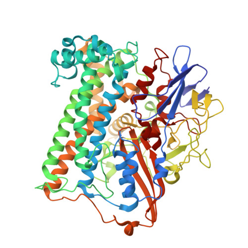
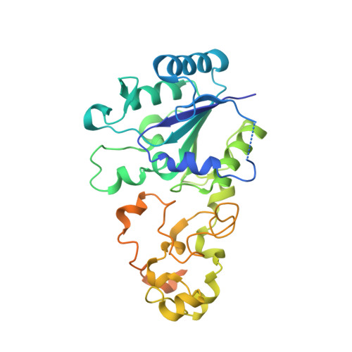
Tracking the route of molecular oxygen in O2-tolerant membrane-bound [NiFe] hydrogenase.

Kalms, J.Schmidt, A.Frielingsdorf, S.Utesch, T.Gotthard, G.von Stetten, D.van der Linden, P.Royant, A.Mroginski, M.A.Carpentier, P.Lenz, O.Scheerer, P.

(2018) Proc Natl Acad Sci U S A 115: E2229-E2237

  • DOI: https://doi.org/10.1073/pnas.1712267115
  • Primary Citation Related Structures: 
    5MDJ, 5MDK, 5MDL

  • PubMed Abstract: 

    [NiFe] hydrogenases catalyze the reversible splitting of H 2 into protons and electrons at a deeply buried active site. The catalytic center can be accessed by gas molecules through a hydrophobic tunnel network. While most [NiFe] hydrogenases are inactivated by O 2 , a small subgroup, including the membrane-bound [NiFe] hydrogenase (MBH) of Ralstonia eutropha , is able to overcome aerobic inactivation by catalytic reduction of O 2 to water. This O 2 tolerance relies on a special [4Fe3S] cluster that is capable of releasing two electrons upon O 2 attack. Here, the O 2 accessibility of the MBH gas tunnel network has been probed experimentally using a "soak-and-freeze" derivatization method, accompanied by protein X-ray crystallography and computational studies. This combined approach revealed several sites of O 2 molecules within a hydrophobic tunnel network leading, via two tunnel entrances, to the catalytic center of MBH. The corresponding site occupancies were related to the O 2 concentrations used for MBH crystal derivatization. The examination of the O 2 -derivatized data furthermore uncovered two unexpected structural alterations at the [4Fe3S] cluster, which might be related to the O 2 tolerance of the enzyme.


  • Organizational Affiliation
    • Charité - Universitätsmedizin Berlin, corporate member of Freie Universität Berlin, Humboldt-Universität zu Berlin, and Berlin Institute of Health, Institute of Medical Physics and Biophysics, Group Protein X-ray Crystallography and Signal Transduction, D-10117 Berlin, Germany.

Macromolecule Content 

  • Total Structure Weight: 104.51 kDa 
  • Atom Count: 7,242 
  • Modeled Residue Count: 859 
  • Deposited Residue Count: 931 
  • Unique protein chains: 2

Macromolecules

Find similar proteins by:|  3D Structure
Entity ID: 1
MoleculeChains  Sequence LengthOrganismDetailsImage
Uptake hydrogenase large subunit; HOXGA [auth L]603Cupriavidus necator H16Mutation(s): 0 
Gene Names: hoxGPHG002
EC: 1.12.99.6
UniProt
Find proteins for P31891 (Cupriavidus necator (strain ATCC 17699 / DSM 428 / KCTC 22496 / NCIMB 10442 / H16 / Stanier 337))
Explore P31891 
Go to UniProtKB:  P31891
Entity Groups
Sequence Clusters30% Identity50% Identity70% Identity90% Identity95% Identity100% Identity
UniProt GroupP31891
Sequence Annotations
Expand
Reference Sequence
Find similar proteins by:|  3D Structure
Entity ID: 2
MoleculeChains  Sequence LengthOrganismDetailsImage
Uptake hydrogenase small subunit; HOXKB [auth S]328Cupriavidus necator H16Mutation(s): 0 
Gene Names: hoxKPHG001
EC: 1.12.99.6
UniProt
Find proteins for P31892 (Cupriavidus necator (strain ATCC 17699 / DSM 428 / KCTC 22496 / NCIMB 10442 / H16 / Stanier 337))
Explore P31892 
Go to UniProtKB:  P31892
Entity Groups
Sequence Clusters30% Identity50% Identity70% Identity90% Identity95% Identity100% Identity
UniProt GroupP31892
Sequence Annotations
Expand
Reference Sequence

Small Molecules

Ligands 6 Unique
IDChains Name / Formula / InChI Key2D Diagram3D Interactions
SF4

Query on SF4


Download:Ideal Coordinates CCD File
E [auth S]IRON/SULFUR CLUSTER
Fe4 S4
LJBDFODJNLIPKO-UHFFFAOYSA-N
F4S

Query on F4S


Download:Ideal Coordinates CCD File
G [auth S]FE4-S3 CLUSTER
Fe4 S3
QQACTBFBZNWJMV-UHFFFAOYSA-N
F3S

Query on F3S


Download:Ideal Coordinates CCD File
F [auth S]FE3-S4 CLUSTER
Fe3 S4
FCXHZBQOKRZXKS-UHFFFAOYSA-N
NFV

Query on NFV


Download:Ideal Coordinates CCD File
C [auth L]NI-FE OXIDIZED ACTIVE CENTER
C3 Fe N2 Ni O2
MPQMGFDSXFFIQL-UHFFFAOYSA-N
CL

Query on CL


Download:Ideal Coordinates CCD File
H [auth S]CHLORIDE ION
Cl
VEXZGXHMUGYJMC-UHFFFAOYSA-M
MG

Query on MG


Download:Ideal Coordinates CCD File
D [auth L]MAGNESIUM ION
Mg
JLVVSXFLKOJNIY-UHFFFAOYSA-N

Experimental Data & Validation

Experimental Data

  • Method: X-RAY DIFFRACTION
  • Resolution: 1.48 Å
  • R-Value Free:  0.172 (Depositor), 0.172 (DCC) 
  • R-Value Work:  0.142 (Depositor), 0.142 (DCC) 
  • R-Value Observed: 0.144 (Depositor) 
Space Group: P 21 21 21
Unit Cell:
Length ( Å )Angle ( ˚ )
a = 73.395α = 90
b = 95.696β = 90
c = 121.565γ = 90
Software Package:
Software NamePurpose
REFMACrefinement
XDSdata reduction
SCALAdata scaling
PHASERphasing

Structure Validation

View Full Validation Report


Entry History 

& Funding Information

Deposition Data


Funding OrganizationLocationGrant Number
German Research FoundationGermanyCluster of Excellence - Unifying Concepts in Catalysis - UniCat - (EXC 314)
German Research FoundationGermanySFB 740 - B6

Revision History  (Full details and data files)

  • Version 1.0: 2018-02-21
    Type: Initial release
  • Version 2.0: 2018-03-28
    Changes: Atomic model, Data collection, Database references, Derived calculations, Structure summary
  • Version 3.0: 2019-01-30
    Changes: Atomic model, Data collection, Derived calculations, Structure summary
  • Version 3.1: 2024-01-17
    Changes: Data collection, Database references, Derived calculations, Refinement description
  • Version 3.2: 2024-11-20
    Changes: Structure summary