5M58 | pdb_00005m58

Crystal structure of CouO, a C-methyltransferase from Streptomyces rishiriensis


Experimental Data Snapshot

  • Method: X-RAY DIFFRACTION
  • Resolution: 2.05 Å
  • R-Value Free: 
    0.264 (Depositor), 0.266 (DCC) 
  • R-Value Work: 
    0.203 (Depositor), 0.211 (DCC) 
  • R-Value Observed: 
    0.207 (Depositor) 

wwPDB Validation 3D Report Full Report

Validation slider image for 5M58

Ligand Structure Quality Assessment 


This is version 1.3 of the entry. See complete history

Literature

Crystal Structure and Catalytic Mechanism of CouO, a Versatile C-Methyltransferase from Streptomyces rishiriensis.

Pavkov-Keller, T.Steiner, K.Faber, M.Tengg, M.Schwab, H.Gruber-Khadjawi, M.Gruber, K.

(2017) PLoS One 12: e0171056-e0171056

  • DOI: https://doi.org/10.1371/journal.pone.0171056
  • Primary Citation Related Structures: 
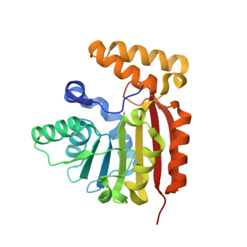
    5M58

  • PubMed Abstract: 

    Friedel-Crafts alkylation of aromatic systems is a classic reaction in organic chemistry, for which regiospecific mono-alkylation, however, is generally difficult to achieve. In nature, methyltransferases catalyze the addition of methyl groups to a wide range of biomolecules thereby modulating the physico-chemical properties of these compounds. Specifically, S-adenosyl-L-methionine dependent C-methyltransferases possess a high potential to serve as biocatalysts in environmentally benign organic syntheses. Here, we report on the high resolution crystal structure of CouO, a C-methyltransferase from Streptomyces rishiriensis involved in the biosynthesis of the antibiotic coumermycin A1. Through molecular docking calculations, site-directed mutagenesis and the comparison with homologous enzymes we identified His120 and Arg121 as key functional residues for the enzymatic activity of this group of C-methyltransferases. The elucidation of the atomic structure and the insight into the catalytic mechanism provide the basis for the (semi)-rational engineering of the enzyme in order to increase the substrate scope as well as to facilitate the acceptance of SAM-analogues as alternative cofactors.


  • Organizational Affiliation
    • Institute of Molecular Biosciences, University of Graz, Graz, Austria.

Macromolecule Content 

  • Total Structure Weight: 52.13 kDa 
  • Atom Count: 3,982 
  • Modeled Residue Count: 459 
  • Deposited Residue Count: 460 
  • Unique protein chains: 1

Macromolecules

Find similar proteins by:|  3D Structure
Entity ID: 1
MoleculeChains  Sequence LengthOrganismDetailsImage
C-methyltransferase CouO
A, B
230Streptomyces rishiriensisMutation(s): 0 
Gene Names: couO
EC: 2.1.1
UniProt
Find proteins for Q9F8T9 (Streptomyces rishiriensis)
Explore Q9F8T9 
Go to UniProtKB:  Q9F8T9
Entity Groups
Sequence Clusters30% Identity50% Identity70% Identity90% Identity95% Identity100% Identity
UniProt GroupQ9F8T9
Sequence Annotations
Expand
Reference Sequence

Experimental Data & Validation

Experimental Data

  • Method: X-RAY DIFFRACTION
  • Resolution: 2.05 Å
  • R-Value Free:  0.264 (Depositor), 0.266 (DCC) 
  • R-Value Work:  0.203 (Depositor), 0.211 (DCC) 
  • R-Value Observed: 0.207 (Depositor) 
Space Group: P 1 21 1
Unit Cell:
Length ( Å )Angle ( ˚ )
a = 33.07α = 90
b = 82.95β = 96.91
c = 76.88γ = 90
Software Package:
Software NamePurpose
REFMACrefinement
MOSFLMdata reduction
SCALAdata scaling
PHASERphasing

Structure Validation

View Full Validation Report


Ligand Structure Quality Assessment 


Entry History 

Deposition Data

Revision History  (Full details and data files)

  • Version 1.0: 2017-02-15
    Type: Initial release
  • Version 1.1: 2019-07-17
    Changes: Data collection
  • Version 1.2: 2024-05-08
    Changes: Data collection, Database references
  • Version 1.3: 2025-12-10
    Changes: Data collection, Structure summary