5M45

Structure of Acetone Carboxylase purified from Xanthobacter autotrophicus


Experimental Data Snapshot

  • Method: X-RAY DIFFRACTION
  • Resolution: 1.87 Å
  • R-Value Free: 0.217 
  • R-Value Work: 0.192 
  • R-Value Observed: 0.193 

wwPDB Validation   3D Report Full Report


Ligand Structure Quality Assessment 


This is version 1.2 of the entry. See complete history


Literature

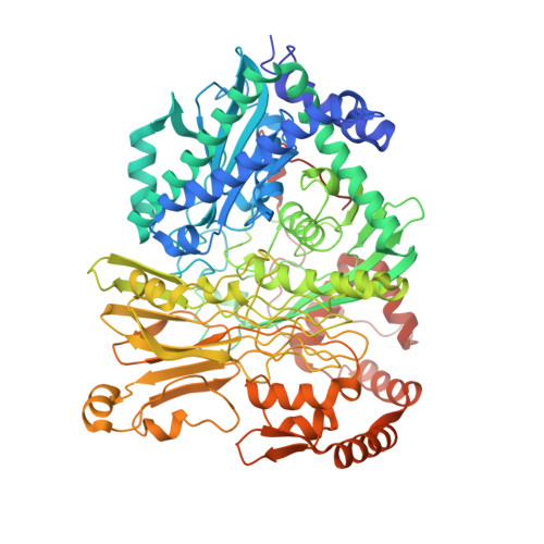
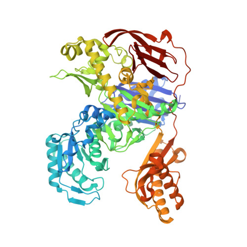
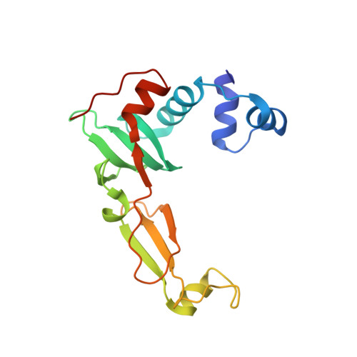
Structural Basis for the Mechanism of ATP-Dependent Acetone Carboxylation.

Mus, F.Eilers, B.J.Alleman, A.B.Kabasakal, B.V.Wells, J.N.Murray, J.W.Nocek, B.P.DuBois, J.L.Peters, J.W.

(2017) Sci Rep 7: 7234-7234

  • DOI: https://doi.org/10.1038/s41598-017-06973-8

  • PubMed Abstract: 

    Microorganisms use carboxylase enzymes to form new carbon-carbon bonds by introducing carbon dioxide gas (CO 2 ) or its hydrated form, bicarbonate (HCO 3 - ), into target molecules. Acetone carboxylases (ACs) catalyze the conversion of substrates acetone and HCO 3 - to form the product acetoacetate. Many bicarbonate-incorporating carboxylases rely on the organic cofactor biotin for the activation of bicarbonate. ACs contain metal ions but not organic cofactors, and use ATP to activate substrates through phosphorylation. How the enzyme coordinates these phosphorylation events and new C-C bond formation in the absence of biotin has remained a mystery since these enzymes were discovered. The first structural rationale for acetone carboxylation is presented here, focusing on the 360 kDa (αβγ) 2 heterohexameric AC from Xanthobacter autotrophicus in the ligand-free, AMP-bound, and acetate coordinated states. These structures suggest successive steps in a catalytic cycle revealing that AC undergoes large conformational changes coupled to substrate activation by ATP to perform C-C bond ligation at a distant Mn center. These results illustrate a new chemical strategy for the conversion of CO 2 into biomass, a process of great significance to the global carbon cycle.


  • Organizational Affiliation

    Insitutite of Biological Chemistry, Washington State University, Pullman, WA, 99164, USA.


Macromolecules
Find similar proteins by:  (by identity cutoff)  |  3D Structure
Entity ID: 1
MoleculeChains Sequence LengthOrganismDetailsImage
Acetone carboxylase alpha subunit
A, D, G, J
776Xanthobacter autotrophicus Py2Mutation(s): 0 
EC: 6.4.1.6
UniProt
Find proteins for Q8RM03 (Xanthobacter autotrophicus (strain ATCC BAA-1158 / Py2))
Explore Q8RM03 
Go to UniProtKB:  Q8RM03
Entity Groups  
Sequence Clusters30% Identity50% Identity70% Identity90% Identity95% Identity100% Identity
UniProt GroupQ8RM03
Sequence Annotations
Expand
  • Reference Sequence
Find similar proteins by:  (by identity cutoff)  |  3D Structure
Entity ID: 2
MoleculeChains Sequence LengthOrganismDetailsImage
Acetone carboxylase beta subunit
B, E, H, K
717Xanthobacter autotrophicus Py2Mutation(s): 0 
EC: 6.4.1.6
UniProt
Find proteins for Q8RM04 (Xanthobacter autotrophicus (strain ATCC BAA-1158 / Py2))
Explore Q8RM04 
Go to UniProtKB:  Q8RM04
Entity Groups  
Sequence Clusters30% Identity50% Identity70% Identity90% Identity95% Identity100% Identity
UniProt GroupQ8RM04
Sequence Annotations
Expand
  • Reference Sequence
Find similar proteins by:  (by identity cutoff)  |  3D Structure
Entity ID: 3
MoleculeChains Sequence LengthOrganismDetailsImage
Acetone carboxylase gamma subunit
C, F, I, L
168Xanthobacter autotrophicus Py2Mutation(s): 0 
EC: 6.4.1.6
UniProt
Find proteins for Q8RM02 (Xanthobacter autotrophicus (strain ATCC BAA-1158 / Py2))
Explore Q8RM02 
Go to UniProtKB:  Q8RM02
Entity Groups  
Sequence Clusters30% Identity50% Identity70% Identity90% Identity95% Identity100% Identity
UniProt GroupQ8RM02
Sequence Annotations
Expand
  • Reference Sequence
Small Molecules
Ligands 6 Unique
IDChains Name / Formula / InChI Key2D Diagram3D Interactions
AMP
Query on AMP

Download Ideal Coordinates CCD File 
FA [auth E],
FB [auth K],
S [auth B],
SA [auth H]
ADENOSINE MONOPHOSPHATE
C10 H14 N5 O7 P
UDMBCSSLTHHNCD-KQYNXXCUSA-N
AE4
Query on AE4

Download Ideal Coordinates CCD File 
CA [auth D],
CB [auth J],
P [auth A],
PA [auth G]
3,6,9,12,15-PENTAOXAHEPTADECAN-1-OL
C12 H26 O6
NJRFAMBTWHGSDE-UHFFFAOYSA-N
ZN
Query on ZN

Download Ideal Coordinates CCD File 
KB [auth L],
LA [auth F],
Y [auth C],
YA [auth I]
ZINC ION
Zn
PTFCDOFLOPIGGS-UHFFFAOYSA-N
ACT
Query on ACT

Download Ideal Coordinates CCD File 
AA [auth D]
AB [auth J]
BA [auth D]
BB [auth J]
JA [auth E]
AA [auth D],
AB [auth J],
BA [auth D],
BB [auth J],
JA [auth E],
JB [auth K],
N [auth A],
NA [auth G],
O [auth A],
OA [auth G],
W [auth B],
XA [auth H]
ACETATE ION
C2 H3 O2
QTBSBXVTEAMEQO-UHFFFAOYSA-M
MN
Query on MN

Download Ideal Coordinates CCD File 
M [auth A],
MA [auth G],
Z [auth D],
ZA [auth J]
MANGANESE (II) ION
Mn
WAEMQWOKJMHJLA-UHFFFAOYSA-N
MG
Query on MG

Download Ideal Coordinates CCD File 
DA [auth D]
DB [auth J]
EA [auth D]
EB [auth J]
GA [auth E]
DA [auth D],
DB [auth J],
EA [auth D],
EB [auth J],
GA [auth E],
GB [auth K],
HA [auth E],
HB [auth K],
IA [auth E],
IB [auth K],
KA [auth E],
LB [auth L],
Q [auth A],
QA [auth G],
R [auth A],
RA [auth G],
T [auth B],
TA [auth H],
U [auth B],
UA [auth H],
V [auth B],
VA [auth H],
WA [auth H],
X [auth B]
MAGNESIUM ION
Mg
JLVVSXFLKOJNIY-UHFFFAOYSA-N
Experimental Data & Validation

Experimental Data

  • Method: X-RAY DIFFRACTION
  • Resolution: 1.87 Å
  • R-Value Free: 0.217 
  • R-Value Work: 0.192 
  • R-Value Observed: 0.193 
  • Space Group: P 1
Unit Cell:
Length ( Å )Angle ( ˚ )
a = 86.915α = 65.89
b = 139.733β = 86.54
c = 165.827γ = 88.69
Software Package:
Software NamePurpose
REFMACrefinement
DIALSdata reduction
xia2data scaling
PHASERphasing

Structure Validation

View Full Validation Report


Ligand Structure Quality Assessment 


Entry History 

Deposition Data

Revision History  (Full details and data files)

  • Version 1.0: 2017-08-09
    Type: Initial release
  • Version 1.1: 2017-08-16
    Changes: Database references
  • Version 1.2: 2024-01-17
    Changes: Data collection, Database references, Derived calculations, Refinement description