5M0H | pdb_00005m0h

Crystal structure of the central flexible region of ASH1 mRNA E3-localization element


Experimental Data Snapshot

  • Method: X-RAY DIFFRACTION
  • Resolution: 2.65 Å
  • R-Value Free: 
    0.240 (Depositor), 0.237 (DCC) 
  • R-Value Work: 
    0.211 (Depositor), 0.216 (DCC) 
  • R-Value Observed: 
    0.212 (Depositor) 

Starting Model: experimental
View more details

wwPDB Validation 3D Report Full Report

Validation slider image for 5M0H

This is version 1.3 of the entry. See complete history

Literature

Molecular architecture and dynamics of ASH1 mRNA recognition by its mRNA-transport complex.

Edelmann, F.T.Schlundt, A.Heym, R.G.Jenner, A.Niedner-Boblenz, A.Syed, M.I.Paillart, J.C.Stehle, R.Janowski, R.Sattler, M.Jansen, R.P.Niessing, D.

(2017) Nat Struct Mol Biol 24: 152-161

  • DOI: https://doi.org/10.1038/nsmb.3351
  • Primary Citation Related Structures: 
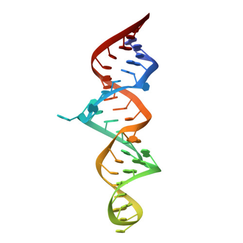
    5M0H, 5M0I, 5M0J

  • PubMed Abstract: 

    mRNA localization is an essential mechanism of gene regulation and is required for processes such as stem-cell division, embryogenesis and neuronal plasticity. It is not known which features in the cis-acting mRNA localization elements (LEs) are specifically recognized by motor-containing transport complexes. To the best of our knowledge, no high-resolution structure is available for any LE in complex with its cognate protein complex. Using X-ray crystallography and complementary techniques, we carried out a detailed assessment of an LE of the ASH1 mRNA from yeast, its complex with its shuttling RNA-binding protein She2p, and its highly specific, cytoplasmic complex with She3p. Although the RNA alone formed a flexible stem loop, She2p binding induced marked conformational changes. However, only joining by the unstructured She3p resulted in specific RNA recognition. The notable RNA rearrangements and joint action of a globular and an unfolded RNA-binding protein offer unprecedented insights into the step-wise maturation of an mRNA-transport complex.


  • Organizational Affiliation
    • Institute of Structural Biology, Helmholtz Zentrum München - German Research Center for Environmental Health, Neuherberg, Germany.

Macromolecule Content 

  • Total Structure Weight: 13.6 kDa 
  • Atom Count: 909 
  • Modeled Residue Count: 42 
  • Deposited Residue Count: 42 
  • Unique nucleic acid chains: 1

Macromolecules

Find similar nucleic acids by:  (by identity cutoff) 
Entity ID: 1
MoleculeChains LengthOrganismImage
ASH1 E3 (42 nt-TL/TLR)42Saccharomyces cerevisiae
Sequence Annotations
Expand
Reference Sequence

Small Molecules

Ligands 1 Unique
IDChains Name / Formula / InChI Key2D Diagram3D Interactions
SO4

Query on SO4


Download:Ideal Coordinates CCD File
B [auth A]SULFATE ION
O4 S
QAOWNCQODCNURD-UHFFFAOYSA-L

Experimental Data & Validation

Experimental Data

  • Method: X-RAY DIFFRACTION
  • Resolution: 2.65 Å
  • R-Value Free:  0.240 (Depositor), 0.237 (DCC) 
  • R-Value Work:  0.211 (Depositor), 0.216 (DCC) 
  • R-Value Observed: 0.212 (Depositor) 
Space Group: P 41 21 2
Unit Cell:
Length ( Å )Angle ( ˚ )
a = 64.32α = 90
b = 64.32β = 90
c = 75.19γ = 90
Software Package:
Software NamePurpose
PHENIXrefinement
XDSdata reduction
XSCALEdata scaling
PHASERphasing

Structure Validation

View Full Validation Report


Entry History 

Deposition Data

Revision History  (Full details and data files)

  • Version 1.0: 2017-01-18
    Type: Initial release
  • Version 1.1: 2017-01-25
    Changes: Database references
  • Version 1.2: 2017-02-15
    Changes: Database references
  • Version 1.3: 2024-01-17
    Changes: Data collection, Database references, Refinement description