5LYG | pdb_00005lyg

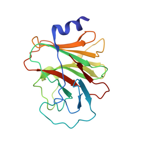
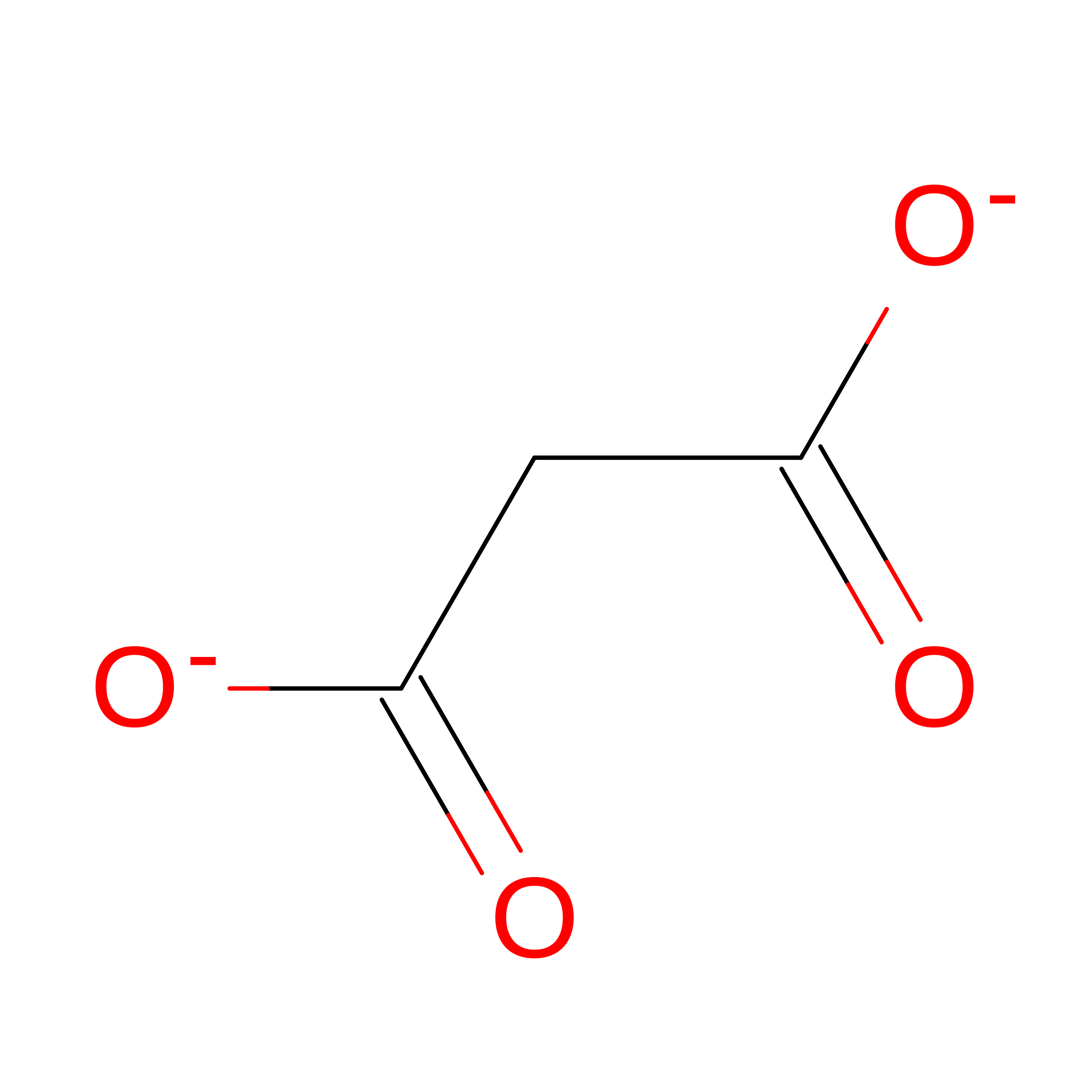
CRYSTAL STRUCTURE OF INTRACELLULAR B30.2 DOMAIN OF BTN3A1 BOUND TO MALONATE


Experimental Data Snapshot

  • Method: X-RAY DIFFRACTION
  • Resolution: 1.60 Å
  • R-Value Free: 
    0.206 (Depositor), 0.205 (DCC) 
  • R-Value Work: 
    0.189 (Depositor), 0.187 (DCC) 
  • R-Value Observed: 
    0.190 (Depositor) 

wwPDB Validation 3D Report Full Report

Validation slider image for 5LYG

This is version 1.2 of the entry. See complete history. 

Literature

BTN3A1 Discriminates gamma delta T Cell Phosphoantigens from Nonantigenic Small Molecules via a Conformational Sensor in Its B30.2 Domain.

Salim, M., Knowles, T.J., Baker, A.T., Davey, M.S., Jeeves, M., Sridhar, P., Wilkie, J., Willcox, C.R., Kadri, H., Taher, T.E., Vantourout, P., Hayday, A., Mehellou, Y., Mohammed, F., Willcox, B.E.

(2017) ACS Chem Biol 12: 2631-2643

  • DOI: https://doi.org/10.1021/acschembio.7b00694
  • Primary Citation Related Structures: 
    5LYG, 5LYK

  • PubMed Abstract: 

    Human Vγ9/Vδ2 T-cells detect tumor cells and microbial infections by recognizing small phosphorylated prenyl metabolites termed phosphoantigens (P-Ag). The type-1 transmembrane protein Butyrophilin 3A1 (BTN3A1) is critical to the P-Ag-mediated activation of Vγ9/Vδ2 T-cells; however, the molecular mechanisms involved in BTN3A1-mediated metabolite sensing are unclear, including how P-Ag's are discriminated from nonantigenic small molecules. Here, we utilized NMR and X-ray crystallography to probe P-Ag sensing by BTN3A1. Whereas the BTN3A1 immunoglobulin variable domain failed to bind P-Ag, the intracellular B30.2 domain bound a range of negatively charged small molecules, including P-Ag, in a positively charged surface pocket. However, NMR chemical shift perturbations indicated BTN3A1 discriminated P-Ag from nonantigenic small molecules by their ability to induce a specific conformational change in the B30.2 domain that propagated from the P-Ag binding site to distal parts of the domain. These results suggest BTN3A1 selectively detects P-Ag intracellularly via a conformational antigenic sensor in its B30.2 domain and have implications for rational design of antigens for Vγ9/Vδ2-based T-cell immunotherapies.


  • Organizational Affiliation: 
    • Institute of Immunology and Immunotherapy, University of Birmingham , Edgbaston, Birmingham, United Kingdom , B15 2TT.

Macromolecule Content 

  • Total Structure Weight: 21.78 kDa 
  • Atom Count: 1,661 
  • Modeled Residue Count: 187 
  • Deposited Residue Count: 187 
  • Unique protein chains: 1

Macromolecules

Find similar proteins by:|  3D Structure
Entity ID: 1
MoleculeChains  Sequence LengthOrganismDetailsImage
Butyrophilin subfamily 3 member A1187Homo sapiensMutation(s): 0 
Gene Names: BTN3A1, BTF5
UniProt & NIH Common Fund Data Resources
Find proteins for O00481 (Homo sapiens)
Explore O00481 
Go to UniProtKB:  O00481
PHAROS:  O00481
GTEx:  ENSG00000026950 
Entity Groups
Sequence Clusters30% Identity50% Identity70% Identity90% Identity95% Identity100% Identity
UniProt GroupO00481
Sequence Annotations
Expand
Reference Sequence

Experimental Data & Validation

Experimental Data

  • Method: X-RAY DIFFRACTION
  • Resolution: 1.60 Å
  • R-Value Free:  0.206 (Depositor), 0.205 (DCC) 
  • R-Value Work:  0.189 (Depositor), 0.187 (DCC) 
  • R-Value Observed: 0.190 (Depositor) 
Space Group: P 21 21 2
Unit Cell:
Length ( Å )Angle ( ˚ )
a = 45.73α = 90
b = 127.22β = 90
c = 39.38γ = 90
Software Package:
Software NamePurpose
REFMACrefinement
XSCALEdata scaling
PDB_EXTRACTdata extraction
XDSdata reduction
MOLREPphasing

Structure Validation

View Full Validation Report


Entry History 

Deposition Data

Revision History  (Full details and data files)

  • Version 1.0: 2017-09-13
    Type: Initial release
  • Version 1.1: 2017-11-01
    Changes: Database references
  • Version 1.2: 2024-05-08
    Changes: Advisory, Data collection, Database references