


How Bacterial Chemoreceptors Evolve Novel Ligand Specificities
Gavira, J.A., Jimenez-Rico, M., Pineda-Molina, E., Krell, T.(2020) mBio
Experimental Data Snapshot
Starting Model: experimental
View more details
wwPDB Validation 3D Report Full Report

(2020) mBio
Macromolecule Content
Entity ID: 1 | |||||
|---|---|---|---|---|---|
| Molecule | Chains | Sequence Length | Organism | Details | Image |
| Chemotactic transducer PctC | 273 | Pseudomonas aeruginosa | Mutation(s): 0 Gene Names: PAMH19_3853 | 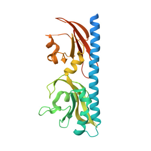 | |
UniProt | |||||
Entity Groups | |||||
| Sequence Clusters | 30% Identity50% Identity70% Identity90% Identity95% Identity100% Identity | ||||
| UniProt Group | Q9HW93 | ||||
Sequence AnnotationsExpand | |||||
Reference Sequence | |||||
| Ligands 4 Unique | |||||
|---|---|---|---|---|---|
| ID | Chains | Name / Formula / InChI Key | 2D Diagram | 3D Interactions | |
| ABU Download:Ideal Coordinates CCD File | H [auth A], M [auth B], P [auth B], W [auth D], Y [auth E] | GAMMA-AMINO-BUTANOIC ACID C4 H9 N O2 BTCSSZJGUNDROE-UHFFFAOYSA-N |  | ||
| SO4 Download:Ideal Coordinates CCD File | AA [auth E] BA [auth E] DA [auth F] FA [auth G] GA [auth G] | SULFATE ION O4 S QAOWNCQODCNURD-UHFFFAOYSA-L |  | ||
| GOL Download:Ideal Coordinates CCD File | EA [auth F], HA [auth G], L [auth A] | GLYCEROL C3 H8 O3 PEDCQBHIVMGVHV-UHFFFAOYSA-N |  | ||
| ACT Download:Ideal Coordinates CCD File | CA [auth F] Q [auth B] R [auth B] S [auth B] T [auth C] | ACETATE ION C2 H3 O2 QTBSBXVTEAMEQO-UHFFFAOYSA-M |  | ||
| Length ( Å ) | Angle ( ˚ ) |
|---|---|
| a = 209.765 | α = 90 |
| b = 209.765 | β = 90 |
| c = 68.885 | γ = 120 |
| Software Name | Purpose |
|---|---|
| SCALA | data scaling |
| SHELX | phasing |
| PHASER | phasing |
| PHENIX | refinement |
| PDB_EXTRACT | data extraction |
| XDS | data reduction |
| Funding Organization | Location | Grant Number |
|---|---|---|
| MICINN | Spain | BIO2013-4297-P |


