5LRW

Structure of Cezanne/OTUD7B OTU domain bound to ubiquitin


Experimental Data Snapshot

  • Method: X-RAY DIFFRACTION
  • Resolution: 2.00 Å
  • R-Value Free: 0.217 
  • R-Value Work: 0.176 
  • R-Value Observed: 0.178 

wwPDB Validation   3D Report Full Report


This is version 2.0 of the entry. See complete history


Literature

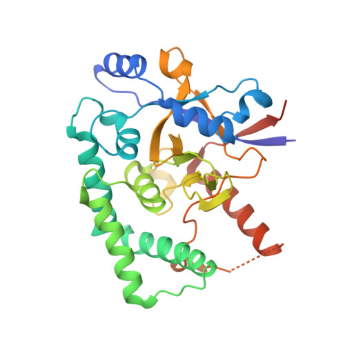
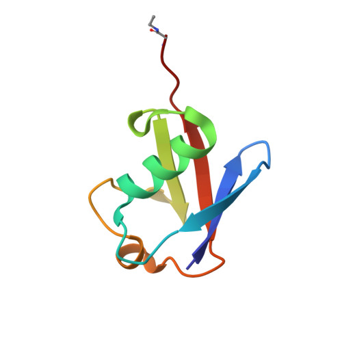
Molecular basis of Lys11-polyubiquitin specificity in the deubiquitinase Cezanne.

Mevissen, T.E.Kulathu, Y.Mulder, M.P.Geurink, P.P.Maslen, S.L.Gersch, M.Elliott, P.R.Burke, J.E.van Tol, B.D.Akutsu, M.El Oualid, F.Kawasaki, M.Freund, S.M.Ovaa, H.Komander, D.

(2016) Nature 538: 402-405

  • DOI: https://doi.org/10.1038/nature19836
  • Primary Citation of Related Structures:  
    5LRU, 5LRV, 5LRW, 5LRX

  • PubMed Abstract: 

    The post-translational modification of proteins with polyubiquitin regulates virtually all aspects of cell biology. Eight distinct chain linkage types co-exist in polyubiquitin and are independently regulated in cells. This 'ubiquitin code' determines the fate of the modified protein. Deubiquitinating enzymes of the ovarian tumour (OTU) family regulate cellular signalling by targeting distinct linkage types within polyubiquitin, and understanding their mechanisms of linkage specificity gives fundamental insights into the ubiquitin system. Here we reveal how the deubiquitinase Cezanne (also known as OTUD7B) specifically targets Lys11-linked polyubiquitin. Crystal structures of Cezanne alone and in complex with monoubiquitin and Lys11-linked diubiquitin, in combination with hydrogen-deuterium exchange mass spectrometry, enable us to reconstruct the enzymatic cycle in great detail. An intricate mechanism of ubiquitin-assisted conformational changes activates the enzyme, and while all chain types interact with the enzymatic S1 site, only Lys11-linked chains can bind productively across the active site and stimulate catalytic turnover. Our work highlights the plasticity of deubiquitinases and indicates that new conformational states can occur when a true substrate, such as diubiquitin, is bound at the active site.


  • Organizational Affiliation

    Medical Research Council Laboratory of Molecular Biology, Francis Crick Avenue, Cambridge, CB2 0QH, UK.


Macromolecules
Find similar proteins by:  (by identity cutoff)  |  3D Structure
Entity ID: 1
MoleculeChains Sequence LengthOrganismDetailsImage
OTU domain-containing protein 7B
A, C
296Homo sapiensMutation(s): 0 
Gene Names: OTUD7BZA20D1
EC: 3.4.19.12
UniProt & NIH Common Fund Data Resources
Find proteins for Q6GQQ9 (Homo sapiens)
Explore Q6GQQ9 
Go to UniProtKB:  Q6GQQ9
PHAROS:  Q6GQQ9
GTEx:  ENSG00000264522 
Entity Groups  
Sequence Clusters30% Identity50% Identity70% Identity90% Identity95% Identity100% Identity
UniProt GroupQ6GQQ9
Sequence Annotations
Expand
  • Reference Sequence
Find similar proteins by:  (by identity cutoff)  |  3D Structure
Entity ID: 2
MoleculeChains Sequence LengthOrganismDetailsImage
Polyubiquitin-B
B, D
76Homo sapiensMutation(s): 0 
Gene Names: UBB
UniProt & NIH Common Fund Data Resources
Find proteins for P0CG47 (Homo sapiens)
Explore P0CG47 
Go to UniProtKB:  P0CG47
PHAROS:  P0CG47
GTEx:  ENSG00000170315 
Entity Groups  
Sequence Clusters30% Identity50% Identity70% Identity90% Identity95% Identity100% Identity
UniProt GroupP0CG47
Sequence Annotations
Expand
  • Reference Sequence
Experimental Data & Validation

Experimental Data

  • Method: X-RAY DIFFRACTION
  • Resolution: 2.00 Å
  • R-Value Free: 0.217 
  • R-Value Work: 0.176 
  • R-Value Observed: 0.178 
  • Space Group: H 3
Unit Cell:
Length ( Å )Angle ( ˚ )
a = 157.56α = 90
b = 157.56β = 90
c = 75.6γ = 120
Software Package:
Software NamePurpose
PHENIXrefinement
XDSdata reduction
Aimlessdata scaling
PHASERphasing

Structure Validation

View Full Validation Report


Entry History & Funding Information

Deposition Data


Funding OrganizationLocationGrant Number
Medical Research Council (United Kingdom)United KingdomU105192732
European Research CouncilUnited Kingdom309756

Revision History  (Full details and data files)

  • Version 1.0: 2016-10-19
    Type: Initial release
  • Version 1.1: 2016-10-26
    Changes: Database references
  • Version 2.0: 2017-09-13
    Changes: Atomic model, Author supporting evidence