5LOI

Crystal structure of Myceliophthora thermophila Rad26 (residues 373-841)


Experimental Data Snapshot

  • Method: X-RAY DIFFRACTION
  • Resolution: 3.15 Å
  • R-Value Free: 0.235 
  • R-Value Work: 0.207 
  • R-Value Observed: 0.210 

wwPDB Validation   3D Report Full Report


This is version 1.2 of the entry. See complete history


Literature

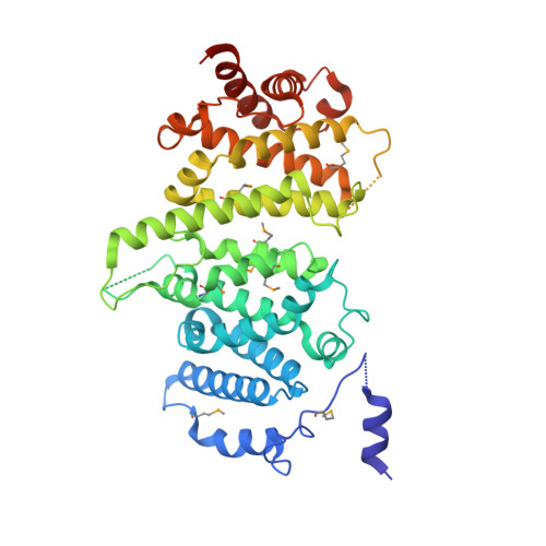
Insights into Rad3 kinase recruitment from the crystal structure of the DNA damage checkpoint protein Rad26.

Andersen, K.R.

(2017) J Biol Chem 292: 8149-8157

  • DOI: https://doi.org/10.1074/jbc.M117.780189
  • Primary Citation of Related Structures:  
    5LOI

  • PubMed Abstract: 

    Metabolic products and environmental factors constantly damage DNA. To protect against these insults and maintain genome integrity, cells have evolved mechanisms to repair DNA lesions. One such mechanism involves Rad3, a master kinase coordinating the DNA damage response. Rad26 is a functional subunit of the Rad3-Rad26 complex and is responsible for bringing the kinase to sites of DNA damage. Here, I present the crystal structure of Rad26 and identify the elements important for recruiting Rad3. The structure suggests that Rad26 is a dimer with a conserved interface in the N-terminal part of the protein. Biochemical data showed that Rad26 uses its C-terminal domain and the flanking kinase-docking motif to bind specific HEAT repeats in Rad3. Analysis of the reconstituted Rad3-Rad26 heterotetrameric complex with electron microscopy enabled me to propose a structural model for its quaternary structure. In conclusion, these results suggest that Rad26 exists as a dimer and provide crucial insight into how Rad3 is recruited and incorporated into the Rad3-Rad26 DNA repair complex.


  • Organizational Affiliation

    Department of Molecular Biology and Genetics, Aarhus University, 8000 Aarhus C, Denmark. Electronic address: kra@mbg.au.dk.


Macromolecules
Find similar proteins by:  (by identity cutoff)  |  3D Structure
Entity ID: 1
MoleculeChains Sequence LengthOrganismDetailsImage
Rad26470Thermothelomyces thermophilus ATCC 42464Mutation(s): 0 
Gene Names: MYCTH_2305697
UniProt
Find proteins for G2QDY6 (Thermothelomyces thermophilus (strain ATCC 42464 / BCRC 31852 / DSM 1799))
Explore G2QDY6 
Go to UniProtKB:  G2QDY6
Entity Groups  
Sequence Clusters30% Identity50% Identity70% Identity90% Identity95% Identity100% Identity
UniProt GroupG2QDY6
Sequence Annotations
Expand
  • Reference Sequence
Small Molecules
Modified Residues  1 Unique
IDChains TypeFormula2D DiagramParent
MSE
Query on MSE
A
L-PEPTIDE LINKINGC5 H11 N O2 SeMET
Experimental Data & Validation

Experimental Data

  • Method: X-RAY DIFFRACTION
  • Resolution: 3.15 Å
  • R-Value Free: 0.235 
  • R-Value Work: 0.207 
  • R-Value Observed: 0.210 
  • Space Group: I 4 2 2
Unit Cell:
Length ( Å )Angle ( ˚ )
a = 210.783α = 90
b = 210.783β = 90
c = 104.766γ = 90
Software Package:
Software NamePurpose
PHENIXrefinement
PDB_EXTRACTdata extraction
XDSdata reduction
SHELXCDphasing

Structure Validation

View Full Validation Report


Entry History 

Deposition Data

Revision History  (Full details and data files)

  • Version 1.0: 2017-03-29
    Type: Initial release
  • Version 1.1: 2017-04-05
    Changes: Database references
  • Version 1.2: 2017-05-31
    Changes: Database references