


Crystal structure of acyl-CoA dehydrogenase (MmgC) from bacillus subtilis.
Baker, G.E., Race, P.R.To be published.
Experimental Data Snapshot
Entity ID: 1 | |||||
|---|---|---|---|---|---|
| Molecule | Chains | Sequence Length | Organism | Details | Image |
| Acyl-CoA dehydrogenase | 379 | Bacillus subtilis subsp. subtilis str. 168 | Mutation(s): 0 Gene Names: mmgC, yqiN, BSU24150 EC: 1.3.99 | 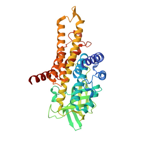 | |
UniProt | |||||
Entity Groups | |||||
| Sequence Clusters | 30% Identity50% Identity70% Identity90% Identity95% Identity100% Identity | ||||
| UniProt Group | P45857 | ||||
Sequence AnnotationsExpand | |||||
Reference Sequence | |||||
| Ligands 2 Unique | |||||
|---|---|---|---|---|---|
| ID | Chains | Name / Formula / InChI Key | 2D Diagram | 3D Interactions | |
| FAD Download:Ideal Coordinates CCD File | AA [auth H] L [auth A] M [auth B] Q [auth C] S [auth D] | FLAVIN-ADENINE DINUCLEOTIDE C27 H33 N9 O15 P2 VWWQXMAJTJZDQX-UYBVJOGSSA-N |  | ||
| GOL Download:Ideal Coordinates CCD File | I [auth A] J [auth A] K [auth A] N [auth C] O [auth C] | GLYCEROL C3 H8 O3 PEDCQBHIVMGVHV-UHFFFAOYSA-N |  | ||
| Length ( Å ) | Angle ( ˚ ) |
|---|---|
| a = 78.168 | α = 90 |
| b = 279.266 | β = 101.4 |
| c = 78.191 | γ = 90 |
| Software Name | Purpose |
|---|---|
| REFMAC | refinement |
| XDS | data reduction |
| Aimless | data scaling |
| PHASER | phasing |


