


Redox-dependent substrate-cofactor interactions in the Michaelis-complex of a flavin-dependent oxidoreductase
Werther, T., Wahlefeld, S., Salewski, J., Kuhlmann, U., Zebger, I., Hildebrandt, P., Dobbek, H.(2017) Nat Commun 8: 16084
Experimental Data Snapshot
Starting Model: experimental
View more details
(2017) Nat Commun 8: 16084
Entity ID: 1 | |||||
|---|---|---|---|---|---|
| Molecule | Chains | Sequence Length | Organism | Details | Image |
| NADH:flavin oxidoreductase | 360 | Pseudomonas putida | Mutation(s): 1 Gene Names: O999_23785 | 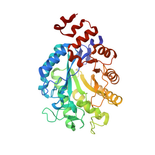 | |
UniProt | |||||
Entity Groups | |||||
| Sequence Clusters | 30% Identity50% Identity70% Identity90% Identity95% Identity100% Identity | ||||
| UniProt Group | A0A1X0ZT96 | ||||
Sequence AnnotationsExpand | |||||
Reference Sequence | |||||
| Ligands 3 Unique | |||||
|---|---|---|---|---|---|
| ID | Chains | Name / Formula / InChI Key | 2D Diagram | 3D Interactions | |
| FNR Download:Ideal Coordinates CCD File | B [auth A] | 1-DEOXY-1-(7,8-DIMETHYL-2,4-DIOXO-3,4-DIHYDRO-2H-BENZO[G]PTERIDIN-1-ID-10(5H)-YL)-5-O-PHOSPHONATO-D-RIBITOL C17 H23 N4 O9 P YTNIXZGTHTVJBW-SCRDCRAPSA-N |  | ||
| 07L Download:Ideal Coordinates CCD File | C [auth A] | 7-hydroxy-2H-chromen-2-one C9 H6 O3 ORHBXUUXSCNDEV-UHFFFAOYSA-N |  | ||
| SO4 Download:Ideal Coordinates CCD File | D [auth A], E [auth A], F [auth A], G [auth A] | SULFATE ION O4 S QAOWNCQODCNURD-UHFFFAOYSA-L |  | ||
| Length ( Å ) | Angle ( ˚ ) |
|---|---|
| a = 57.62 | α = 90 |
| b = 83.46 | β = 90 |
| c = 156.87 | γ = 90 |
| Software Name | Purpose |
|---|---|
| PHENIX | refinement |
| XSCALE | data scaling |
| PHASER | phasing |
| PDB_EXTRACT | data extraction |
| XDS | data reduction |


