


Crystal structure of mouse CARM1 in complex with ligands
Cura, V., Marechal, N., Mailliot, J., Troffer-Charlier, N., Hassenboehler, P., Wurtz, J.M., Bonnefond, L., Cavarelli, J.To be published.
Experimental Data Snapshot
Starting Model: experimental
View more details
Entity ID: 1 | |||||
|---|---|---|---|---|---|
| Molecule | Chains | Sequence Length | Organism | Details | Image |
| Histone-arginine methyltransferase CARM1 | 371 | Mus musculus | Mutation(s): 0 Gene Names: Carm1, Prmt4 EC: 2.1.1.319 | 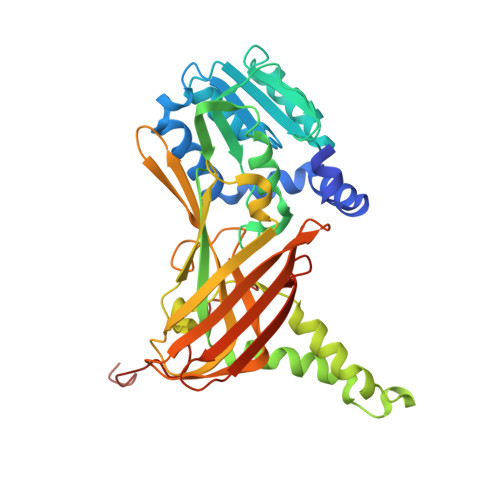 | |
UniProt & NIH Common Fund Data Resources | |||||
IMPC: MGI:1913208 | |||||
Entity Groups | |||||
| Sequence Clusters | 30% Identity50% Identity70% Identity90% Identity95% Identity100% Identity | ||||
| UniProt Group | Q9WVG6 | ||||
Sequence AnnotationsExpand | |||||
Reference Sequence | |||||
| Ligands 5 Unique | |||||
|---|---|---|---|---|---|
| ID | Chains | Name / Formula / InChI Key | 2D Diagram | 3D Interactions | |
| 6YB Download:Ideal Coordinates CCD File | E [auth A], N [auth B], U [auth C], Y [auth D] | (2~{S})-4-[[(2~{R},3~{S},4~{R},5~{R})-5-(6-aminopurin-9-yl)-3,4-bis(oxidanyl)oxolan-2-yl]methyl-(2-carbamimidamidoethyl)amino]-2-azanyl-butanoic acid C17 H28 N10 O5 JNOBHECZYPSIIL-OPYVMVOTSA-N |  | ||
| P33 Download:Ideal Coordinates CCD File | R [auth B] | 3,6,9,12,15,18-HEXAOXAICOSANE-1,20-DIOL C14 H30 O8 XPJRQAIZZQMSCM-UHFFFAOYSA-N |  | ||
| PEG Download:Ideal Coordinates CCD File | M [auth A], W [auth C], X [auth C] | DI(HYDROXYETHYL)ETHER C4 H10 O3 MTHSVFCYNBDYFN-UHFFFAOYSA-N |  | ||
| DXE Download:Ideal Coordinates CCD File | J [auth A], K [auth A], L [auth A], S [auth B], T [auth B] | 1,2-DIMETHOXYETHANE C4 H10 O2 XTHFKEDIFFGKHM-UHFFFAOYSA-N |  | ||
| EDO Download:Ideal Coordinates CCD File | F [auth A] G [auth A] H [auth A] I [auth A] O [auth B] | 1,2-ETHANEDIOL C2 H6 O2 LYCAIKOWRPUZTN-UHFFFAOYSA-N |  | ||
| Length ( Å ) | Angle ( ˚ ) |
|---|---|
| a = 74.637 | α = 90 |
| b = 98.161 | β = 90 |
| c = 206.499 | γ = 90 |
| Software Name | Purpose |
|---|---|
| PHENIX | refinement |
| HKL-2000 | data reduction |
| SCALEPACK | data scaling |
| PHASER | phasing |


