5LIH

Structure of a peptide-substrate bound to PKCiota core kinase domain


Experimental Data Snapshot

  • Method: X-RAY DIFFRACTION
  • Resolution: 3.25 Å
  • R-Value Free: 0.284 
  • R-Value Work: 0.257 
  • R-Value Observed: 0.258 

wwPDB Validation   3D Report Full Report


Ligand Structure Quality Assessment 


This is version 1.2 of the entry. See complete history


Literature

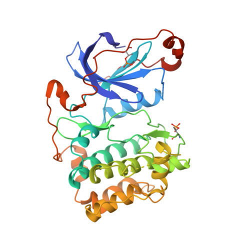
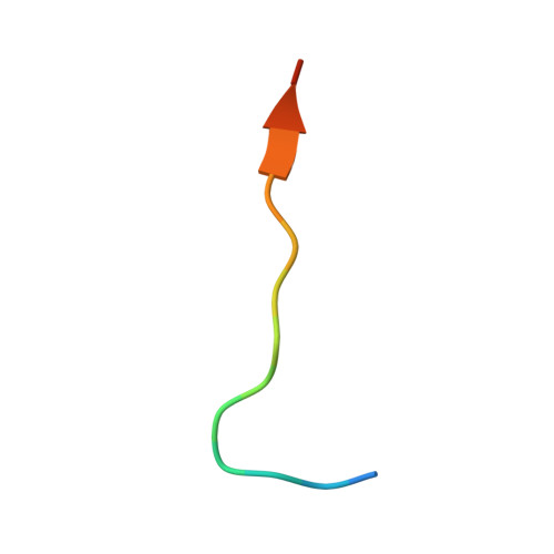
aPKC Inhibition by Par3 CR3 Flanking Regions Controls Substrate Access and Underpins Apical-Junctional Polarization.

Soriano, E.V.Ivanova, M.E.Fletcher, G.Riou, P.Knowles, P.P.Barnouin, K.Purkiss, A.Kostelecky, B.Saiu, P.Linch, M.Elbediwy, A.Kjr, S.O'Reilly, N.Snijders, A.P.Parker, P.J.Thompson, B.J.McDonald, N.Q.

(2016) Dev Cell 38: 384-398

  • DOI: https://doi.org/10.1016/j.devcel.2016.07.018
  • Primary Citation of Related Structures:  
    5LI1, 5LI9, 5LIH

  • PubMed Abstract: 

    Atypical protein kinase C (aPKC) is a key apical-basal polarity determinant and Par complex component. It is recruited by Par3/Baz (Bazooka in Drosophila) into epithelial apical domains through high-affinity interaction. Paradoxically, aPKC also phosphorylates Par3/Baz, provoking its relocalization to adherens junctions (AJs). We show that Par3 conserved region 3 (CR3) forms a tight inhibitory complex with a primed aPKC kinase domain, blocking substrate access. A CR3 motif flanking its PKC consensus site disrupts the aPKC kinase N lobe, separating P-loop/αB/αC contacts. A second CR3 motif provides a high-affinity anchor. Mutation of either motif switches CR3 to an efficient in vitro substrate by exposing its phospho-acceptor site. In vivo, mutation of either CR3 motif alters Par3/Baz localization from apical to AJs. Our results reveal how Par3/Baz CR3 can antagonize aPKC in stable apical Par complexes and suggests that modulation of CR3 inhibitory arms or opposing aPKC pockets would perturb the interaction, promoting Par3/Baz phosphorylation.


  • Organizational Affiliation

    Structural Biology, The Francis Crick Institute, 44 Lincoln's Inn Fields, London WC2A 3LY, UK.


Macromolecules
Find similar proteins by:  (by identity cutoff)  |  3D Structure
Entity ID: 1
MoleculeChains Sequence LengthOrganismDetailsImage
Protein kinase C iota type
A, B
349Homo sapiensMutation(s): 0 
Gene Names: PRKCIDXS1179E
EC: 2.7.11.13
UniProt & NIH Common Fund Data Resources
Find proteins for P41743 (Homo sapiens)
Explore P41743 
Go to UniProtKB:  P41743
PHAROS:  P41743
GTEx:  ENSG00000163558 
Entity Groups  
Sequence Clusters30% Identity50% Identity70% Identity90% Identity95% Identity100% Identity
UniProt GroupP41743
Sequence Annotations
Expand
  • Reference Sequence

Find similar proteins by:  Sequence   |   3D Structure  

Entity ID: 2
MoleculeChains Sequence LengthOrganismDetailsImage
PKC Epsilon pseudo substrate sequenceC [auth F],
D [auth G]
16Homo sapiensMutation(s): 0 
UniProt & NIH Common Fund Data Resources
Find proteins for Q02156 (Homo sapiens)
Explore Q02156 
Go to UniProtKB:  Q02156
PHAROS:  Q02156
GTEx:  ENSG00000171132 
Entity Groups  
Sequence Clusters30% Identity50% Identity70% Identity90% Identity95% Identity100% Identity
UniProt GroupQ02156
Sequence Annotations
Expand
  • Reference Sequence
Small Molecules
Ligands 4 Unique
IDChains Name / Formula / InChI Key2D Diagram3D Interactions
ADP
Query on ADP

Download Ideal Coordinates CCD File 
E [auth A],
L [auth B]
ADENOSINE-5'-DIPHOSPHATE
C10 H15 N5 O10 P2
XTWYTFMLZFPYCI-KQYNXXCUSA-N
AF3
Query on AF3

Download Ideal Coordinates CCD File 
F [auth A],
I [auth A],
M [auth B],
P [auth B]
ALUMINUM FLUORIDE
Al F3
KLZUFWVZNOTSEM-UHFFFAOYSA-K
SCN
Query on SCN

Download Ideal Coordinates CCD File 
J [auth A],
Q [auth B]
THIOCYANATE ION
C N S
ZMZDMBWJUHKJPS-UHFFFAOYSA-M
MN
Query on MN

Download Ideal Coordinates CCD File 
G [auth A],
H [auth A],
K [auth A],
N [auth B],
O [auth B]
MANGANESE (II) ION
Mn
WAEMQWOKJMHJLA-UHFFFAOYSA-N
Modified Residues  1 Unique
IDChains TypeFormula2D DiagramParent
TPO
Query on TPO
A, B
L-PEPTIDE LINKINGC4 H10 N O6 PTHR
Experimental Data & Validation

Experimental Data

  • Method: X-RAY DIFFRACTION
  • Resolution: 3.25 Å
  • R-Value Free: 0.284 
  • R-Value Work: 0.257 
  • R-Value Observed: 0.258 
  • Space Group: P 21 21 21
Unit Cell:
Length ( Å )Angle ( ˚ )
a = 78.98α = 90
b = 84.23β = 90
c = 111.83γ = 90
Software Package:
Software NamePurpose
PHENIXrefinement
xia2data reduction
SCALAdata scaling
PHASERphasing

Structure Validation

View Full Validation Report


Ligand Structure Quality Assessment 


Entry History 

Deposition Data

Revision History  (Full details and data files)

  • Version 1.0: 2016-09-14
    Type: Initial release
  • Version 1.1: 2018-10-17
    Changes: Data collection, Source and taxonomy, Structure summary
  • Version 1.2: 2024-01-10
    Changes: Data collection, Database references, Derived calculations, Refinement description, Structure summary