5LGH | pdb_00005lgh

Afamin antibody fragment, N14 Fab, L1- glycosilated, crystal form II, same as 5L7X, but isomorphous setting indexed same as 5L88, 5L9D


Experimental Data Snapshot

  • Method: X-RAY DIFFRACTION
  • Resolution: 1.86 Å
  • R-Value Free: 
    0.228 (Depositor), 0.218 (DCC) 
  • R-Value Work: 
    0.190 (Depositor) 
  • R-Value Observed: 
    0.192 (Depositor) 

Starting Model: experimental
View more details

wwPDB Validation 3D Report Full Report

Validation slider image for 5LGH

Ligand Structure Quality Assessment 


This is version 1.5 of the entry. See complete history

Literature

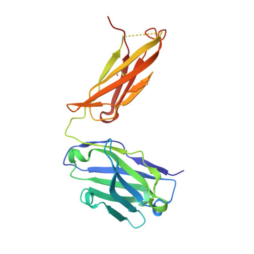
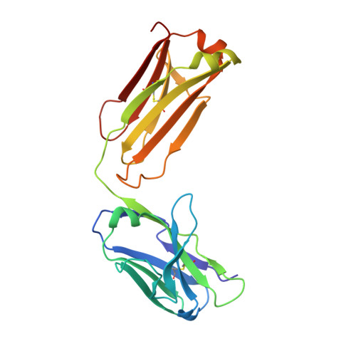
The N14 anti-afamin antibody Fab: a rare VL1 CDR glycosylation, crystallographic re-sequencing, molecular plasticity and conservative versus enthusiastic modelling.

Naschberger, A.Furnrohr, B.G.Lenac Rovis, T.Malic, S.Scheffzek, K.Dieplinger, H.Rupp, B.

(2016) Acta Crystallogr D Struct Biol 72: 1267-1280

  • DOI: https://doi.org/10.1107/S205979831601723X
  • Primary Citation Related Structures: 
    5L7X, 5L88, 5L9D, 5LGH

  • PubMed Abstract: 

    The monoclonal antibody N14 is used as a detection antibody in ELISA kits for the human glycoprotein afamin, a member of the albumin family, which has recently gained interest in the capture and stabilization of Wnt signalling proteins, and for its role in metabolic syndrome and papillary thyroid carcinoma. As a rare occurrence, the N14 Fab is N-glycosylated at Asn26L at the onset of the V L 1 antigen-binding loop, with the α-1-6 core fucosylated complex glycan facing out of the L1 complementarity-determining region. The crystal structures of two non-apparent (pseudo) isomorphous crystals of the N14 Fab were analyzed, which differ significantly in the elbow angles, thereby cautioning against the overinterpretation of domain movements upon antigen binding. In addition, the map quality at 1.9 Å resolution was sufficient to crystallographically re-sequence the variable V L and V H domains and to detect discrepancies in the hybridoma-derived sequence. Finally, a conservatively refined parsimonious model is presented and its statistics are compared with those from a less conservatively built model that has been modelled more enthusiastically. Improvements to the PDB validation reports affecting ligands, clashscore and buried surface calculations are suggested.


  • Organizational Affiliation
    • Division of Biological Chemistry, Medical University of Innsbruck, Innrain 80, 6020 Innsbruck, Austria.

Macromolecule Content 

  • Total Structure Weight: 48.32 kDa 
  • Atom Count: 3,555 
  • Modeled Residue Count: 419 
  • Deposited Residue Count: 435 
  • Unique protein chains: 2

Macromolecules

Find similar proteins by:|  3D Structure
Entity ID: 1
MoleculeChains  Sequence LengthOrganismDetailsImage
MOUSE ANTIBODY FAB FRAGMENT, IGG1-KAPPA HEAVY CHAINA [auth H]220Mus musculusMutation(s): 0 
Find similar proteins by:|  3D Structure
Entity ID: 2
MoleculeChains  Sequence LengthOrganismDetailsImage
MOUSE ANTIBODY FAB FRAGMENT, IGG1-KAPPA LIGHT CHAINB [auth L]215Mus musculusMutation(s): 0 

Small Molecules

Ligands 4 Unique
IDChains Name / Formula / InChI Key2D Diagram3D Interactions
1PE

Query on 1PE


Download:Ideal Coordinates CCD File
G [auth L]PENTAETHYLENE GLYCOL
C10 H22 O6
JLFNLZLINWHATN-UHFFFAOYSA-N
NAG

Query on NAG


Download:Ideal Coordinates CCD File
H [auth L]2-acetamido-2-deoxy-beta-D-glucopyranose
C8 H15 N O6
OVRNDRQMDRJTHS-FMDGEEDCSA-N
PG4

Query on PG4


Download:Ideal Coordinates CCD File
C [auth H]TETRAETHYLENE GLYCOL
C8 H18 O5
UWHCKJMYHZGTIT-UHFFFAOYSA-N
PEG

Query on PEG


Download:Ideal Coordinates CCD File
D [auth H],
E [auth H],
F [auth H]
DI(HYDROXYETHYL)ETHER
C4 H10 O3
MTHSVFCYNBDYFN-UHFFFAOYSA-N

Experimental Data & Validation

Experimental Data

  • Method: X-RAY DIFFRACTION
  • Resolution: 1.86 Å
  • R-Value Free:  0.228 (Depositor), 0.218 (DCC) 
  • R-Value Work:  0.190 (Depositor) 
  • R-Value Observed: 0.192 (Depositor) 
Space Group: P 21 21 21
Unit Cell:
Length ( Å )Angle ( ˚ )
a = 72.196α = 90
b = 67.491β = 90
c = 88.941γ = 90
Software Package:
Software NamePurpose
REFMACrefinement
XDSdata reduction
autoPROCdata reduction
XSCALEdata scaling
Aimlessdata scaling
BALBESphasing

Structure Validation

View Full Validation Report


Ligand Structure Quality Assessment 


Entry History 

Deposition Data

Revision History  (Full details and data files)

  • Version 1.0: 2016-08-03
    Type: Initial release
  • Version 1.1: 2016-12-07
    Changes: Database references
  • Version 1.2: 2016-12-21
    Changes: Database references
  • Version 1.3: 2020-07-29
    Type: Remediation
    Reason: Carbohydrate remediation
    Changes: Data collection, Derived calculations, Refinement description, Structure summary
  • Version 1.4: 2024-01-10
    Changes: Data collection, Database references, Refinement description, Structure summary
  • Version 1.5: 2024-11-13
    Changes: Structure summary