5LBO

Crystal structure of human phosphodiesterase 4D2 catalytic domain with inhibitor NPD-001


Experimental Data Snapshot

  • Method: X-RAY DIFFRACTION
  • Resolution: 2.25 Å
  • R-Value Free: 0.210 
  • R-Value Work: 0.174 
  • R-Value Observed: 0.175 

wwPDB Validation   3D Report Full Report


Ligand Structure Quality Assessment 


This is version 1.3 of the entry. See complete history


Literature

Targeting a Subpocket in Trypanosoma brucei Phosphodiesterase B1 (TbrPDEB1) Enables the Structure-Based Discovery of Selective Inhibitors with Trypanocidal Activity.

Blaazer, A.R.Singh, A.K.de Heuvel, E.Edink, E.Orrling, K.M.Veerman, J.J.N.van den Bergh, T.Jansen, C.Balasubramaniam, E.Mooij, W.J.Custers, H.Sijm, M.Tagoe, D.N.A.Kalejaiye, T.D.Munday, J.C.Tenor, H.Matheeussen, A.Wijtmans, M.Siderius, M.de Graaf, C.Maes, L.de Koning, H.P.Bailey, D.S.Sterk, G.J.de Esch, I.J.P.Brown, D.G.Leurs, R.

(2018) J Med Chem 61: 3870-3888

  • DOI: https://doi.org/10.1021/acs.jmedchem.7b01670
  • Primary Citation of Related Structures:  
    5G2B, 5G57, 5G5V, 5L8C, 5L8Y, 5L9H, 5LAQ, 5LBO

  • PubMed Abstract: 

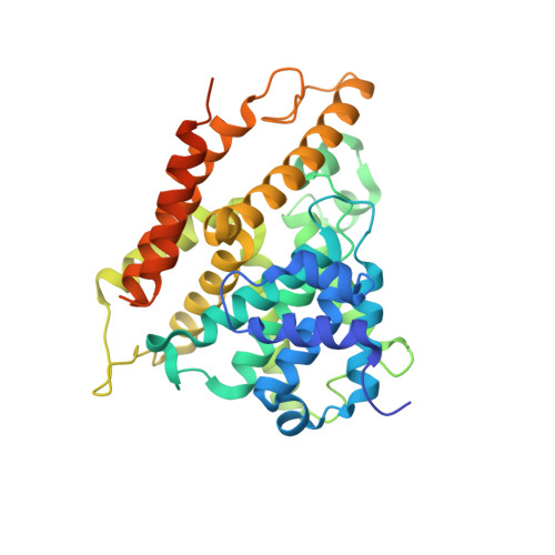
    Several trypanosomatid cyclic nucleotide phosphodiesterases (PDEs) possess a unique, parasite-specific cavity near the ligand-binding region that is referred to as the P-pocket. One of these enzymes, Trypanosoma brucei PDE B1 (TbrPDEB1), is considered a drug target for the treatment of African sleeping sickness. Here, we elucidate the molecular determinants of inhibitor binding and reveal that the P-pocket is amenable to directed design. By iterative cycles of design, synthesis, and pharmacological evaluation and by elucidating the structures of inhibitor-bound TbrPDEB1, hPDE4B, and hPDE4D complexes, we have developed 4a,5,8,8a-tetrahydrophthalazinones as the first selective TbrPDEB1 inhibitor series. Two of these, 8 (NPD-008) and 9 (NPD-039), were potent ( K i = 100 nM) TbrPDEB1 inhibitors with antitrypanosomal effects (IC 50 = 5.5 and 6.7 μM, respectively). Treatment of parasites with 8 caused an increase in intracellular cyclic adenosine monophosphate (cAMP) levels and severe disruption of T. brucei cellular organization, chemically validating trypanosomal PDEs as therapeutic targets in trypanosomiasis.


  • Organizational Affiliation

    Division of Medicinal Chemistry, Amsterdam Institute for Molecules, Medicines and Systems , Vrije Universiteit Amsterdam , 1081 HZ Amsterdam , The Netherlands.


Macromolecules
Find similar proteins by:  (by identity cutoff)  |  3D Structure
Entity ID: 1
MoleculeChains Sequence LengthOrganismDetailsImage
cAMP-specific 3',5'-cyclic phosphodiesterase 4D
A, B, C, D
364Homo sapiensMutation(s): 0 
Gene Names: PDE4DDPDE3
EC: 3.1.4.53
UniProt & NIH Common Fund Data Resources
Find proteins for Q08499 (Homo sapiens)
Explore Q08499 
Go to UniProtKB:  Q08499
PHAROS:  Q08499
GTEx:  ENSG00000113448 
Entity Groups  
Sequence Clusters30% Identity50% Identity70% Identity90% Identity95% Identity100% Identity
UniProt GroupQ08499
Sequence Annotations
Expand
  • Reference Sequence
Small Molecules
Ligands 7 Unique
IDChains Name / Formula / InChI Key2D Diagram3D Interactions
6M5
Query on 6M5

Download Ideal Coordinates CCD File 
BA [auth B],
JA [auth C],
O [auth A],
YA [auth D]
(4~{a}~{S},8~{a}~{R})-2-cycloheptyl-4-[4-methoxy-3-[4-[4-(1~{H}-1,2,3,4-tetrazol-5-yl)phenoxy]butoxy]phenyl]-4~{a},5,8,8~{a}-tetrahydrophthalazin-1-one
C33 H40 N6 O4
DNDNLFXKQSTINI-WUFINQPMSA-N
EPE
Query on EPE

Download Ideal Coordinates CCD File 
AA [auth B],
IA [auth C]
4-(2-HYDROXYETHYL)-1-PIPERAZINE ETHANESULFONIC ACID
C8 H18 N2 O4 S
JKMHFZQWWAIEOD-UHFFFAOYSA-N
DTT
Query on DTT

Download Ideal Coordinates CCD File 
N [auth A],
Q [auth A],
XA [auth D]
2,3-DIHYDROXY-1,4-DITHIOBUTANE
C4 H10 O2 S2
VHJLVAABSRFDPM-IMJSIDKUSA-N
PEG
Query on PEG

Download Ideal Coordinates CCD File 
VA [auth D],
WA [auth D]
DI(HYDROXYETHYL)ETHER
C4 H10 O3
MTHSVFCYNBDYFN-UHFFFAOYSA-N
ZN
Query on ZN

Download Ideal Coordinates CCD File 
CA [auth C],
E [auth A],
KA [auth D],
R [auth B]
ZINC ION
Zn
PTFCDOFLOPIGGS-UHFFFAOYSA-N
EDO
Query on EDO

Download Ideal Coordinates CCD File 
EA [auth C]
FA [auth C]
G [auth A]
GA [auth C]
H [auth A]
EA [auth C],
FA [auth C],
G [auth A],
GA [auth C],
H [auth A],
HA [auth C],
I [auth A],
J [auth A],
K [auth A],
L [auth A],
M [auth A],
MA [auth D],
NA [auth D],
OA [auth D],
P [auth A],
PA [auth D],
QA [auth D],
RA [auth D],
SA [auth D],
T [auth B],
TA [auth D],
U [auth B],
UA [auth D],
V [auth B],
W [auth B],
X [auth B],
Y [auth B],
Z [auth B]
1,2-ETHANEDIOL
C2 H6 O2
LYCAIKOWRPUZTN-UHFFFAOYSA-N
MG
Query on MG

Download Ideal Coordinates CCD File 
DA [auth C],
F [auth A],
LA [auth D],
S [auth B]
MAGNESIUM ION
Mg
JLVVSXFLKOJNIY-UHFFFAOYSA-N
Binding Affinity Annotations 
IDSourceBinding Affinity
6M5 BindingDB:  5LBO IC50: 0.63 (nM) from 1 assay(s)
Experimental Data & Validation

Experimental Data

  • Method: X-RAY DIFFRACTION
  • Resolution: 2.25 Å
  • R-Value Free: 0.210 
  • R-Value Work: 0.174 
  • R-Value Observed: 0.175 
  • Space Group: P 21 21 21
Unit Cell:
Length ( Å )Angle ( ˚ )
a = 98.223α = 90
b = 111.092β = 90
c = 160.798γ = 90
Software Package:
Software NamePurpose
REFMACrefinement
XDSdata reduction
Aimlessdata scaling
PHASERphasing

Structure Validation

View Full Validation Report


Ligand Structure Quality Assessment 


Entry History 

Deposition Data

Revision History  (Full details and data files)

  • Version 1.0: 2018-03-14
    Type: Initial release
  • Version 1.1: 2018-05-02
    Changes: Data collection, Database references
  • Version 1.2: 2018-05-23
    Changes: Data collection, Database references
  • Version 1.3: 2024-01-10
    Changes: Data collection, Database references, Derived calculations, Refinement description