5LBA | pdb_00005lba

Crystal structure of human RECQL5 helicase in complex with DSPL fragment(1-cyclohexyl-3-(oxolan-2-ylmethyl)urea, SGC - Diamond XChem I04-1 fragment screening.


Experimental Data Snapshot

  • Method: X-RAY DIFFRACTION
  • Resolution: 2.50 Å
  • R-Value Free: 
    0.303 (Depositor), 0.331 (DCC) 
  • R-Value Work: 
    0.237 (Depositor), 0.271 (DCC) 
  • R-Value Observed: 
    0.240 (Depositor) 

Starting Model: experimental
View more details

wwPDB Validation 3D Report Full Report

Validation slider image for 5LBA

Ligand Structure Quality Assessment 


This is version 1.3 of the entry. See complete history

Literature

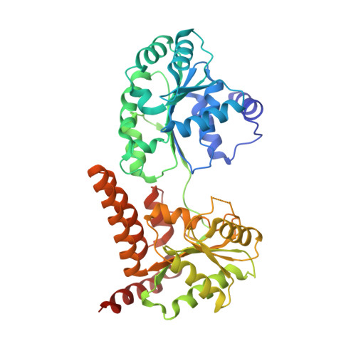
Crystal structure of human RECQL5 helicase in complex with 3D fragment (1-cyclohexyl-3-(oxolan-2-ylmethyl)urea)

Newman, J.A.Talon, R.Aitkenhead, H.Savitsky, P.Krojer, T.von Delft, F.Arrowsmith, C.H.Edwards, A.M.Bountra, C.Gileadi, O.

To be published.

Macromolecule Content 

  • Total Structure Weight: 200.68 kDa 
  • Atom Count: 13,954 
  • Modeled Residue Count: 1,733 
  • Deposited Residue Count: 1,780 
  • Unique protein chains: 1

Macromolecules

Find similar proteins by:|  3D Structure
Entity ID: 1
MoleculeChains  Sequence LengthOrganismDetailsImage
ATP-dependent DNA helicase Q5A [auth B],
B [auth A],
C,
D
445Homo sapiensMutation(s): 0 
Gene Names: RECQL5RECQ5
EC: 3.6.4.12 (PDB Primary Data), 5.6.2.4 (UniProt)
UniProt & NIH Common Fund Data Resources
Find proteins for O94762 (Homo sapiens)
Explore O94762 
Go to UniProtKB:  O94762
PHAROS:  O94762
GTEx:  ENSG00000108469 
Entity Groups
Sequence Clusters30% Identity50% Identity70% Identity90% Identity95% Identity100% Identity
UniProt GroupO94762
Sequence Annotations
Expand
Reference Sequence

Small Molecules

Ligands 5 Unique
IDChains Name / Formula / InChI Key2D Diagram3D Interactions
ADP

Query on ADP


Download:Ideal Coordinates CCD File
G [auth B],
K [auth A],
P [auth C],
S [auth D]
ADENOSINE-5'-DIPHOSPHATE
C10 H15 N5 O10 P2
XTWYTFMLZFPYCI-KQYNXXCUSA-N
6SV

Query on 6SV


Download:Ideal Coordinates CCD File
I [auth B]1-cyclohexyl-3-[[(2~{R})-oxolan-2-yl]methyl]urea
C12 H22 N2 O2
RYUZWZOTJULRNS-LLVKDONJSA-N
DMS

Query on DMS


Download:Ideal Coordinates CCD File
H [auth B],
M [auth A],
T [auth D]
DIMETHYL SULFOXIDE
C2 H6 O S
IAZDPXIOMUYVGZ-UHFFFAOYSA-N
ZN

Query on ZN


Download:Ideal Coordinates CCD File
F [auth B],
L [auth A],
O [auth C],
R [auth D]
ZINC ION
Zn
PTFCDOFLOPIGGS-UHFFFAOYSA-N
MG

Query on MG


Download:Ideal Coordinates CCD File
E [auth B],
J [auth A],
N [auth C],
Q [auth D]
MAGNESIUM ION
Mg
JLVVSXFLKOJNIY-UHFFFAOYSA-N

Experimental Data & Validation

Experimental Data

  • Method: X-RAY DIFFRACTION
  • Resolution: 2.50 Å
  • R-Value Free:  0.303 (Depositor), 0.331 (DCC) 
  • R-Value Work:  0.237 (Depositor), 0.271 (DCC) 
  • R-Value Observed: 0.240 (Depositor) 
Space Group: P 1
Unit Cell:
Length ( Å )Angle ( ˚ )
a = 63.926α = 109.9
b = 85.261β = 90.02
c = 105.915γ = 97.01
Software Package:
Software NamePurpose
REFMACrefinement
XDSdata reduction
XDSdata scaling
REFMACphasing

Structure Validation

View Full Validation Report


Ligand Structure Quality Assessment 


Entry History 

& Funding Information

Deposition Data


Funding OrganizationLocationGrant Number
Wellcome TrustUnited Kingdom--

Revision History  (Full details and data files)

  • Version 1.0: 2016-07-06
    Type: Initial release
  • Version 1.1: 2018-01-24
    Changes: Database references, Structure summary
  • Version 1.2: 2024-01-10
    Changes: Data collection, Database references, Derived calculations, Refinement description
  • Version 1.3: 2024-10-16
    Changes: Structure summary