5L0B | pdb_00005l0b

Crystal Structure of Autotaxin and Compound 1


Experimental Data Snapshot

  • Method: X-RAY DIFFRACTION
  • Resolution: 2.41 Å
  • R-Value Free: 
    0.209 (Depositor), 0.223 (DCC) 
  • R-Value Work: 
    0.176 (Depositor), 0.186 (DCC) 
  • R-Value Observed: 
    0.176 (Depositor) 

wwPDB Validation 3D Report Full Report

Validation slider image for 5L0B

Ligand Structure Quality Assessment 


This is version 2.1 of the entry. See complete history

Literature

Novel Autotaxin Inhibitors for the Treatment of Osteoarthritis Pain: Lead Optimization via Structure-Based Drug Design.

Jones, S.B.Pfeifer, L.A.Bleisch, T.J.Beauchamp, T.J.Durbin, J.D.Klimkowski, V.J.Hughes, N.E.Rito, C.J.Dao, Y.Gruber, J.M.Bui, H.Chambers, M.G.Chandrasekhar, S.Lin, C.McCann, D.J.Mudra, D.R.Oskins, J.L.Swearingen, C.A.Thirunavukkarasu, K.Norman, B.H.

(2016) ACS Med Chem Lett 7: 857-861

  • DOI: https://doi.org/10.1021/acsmedchemlett.6b00207
  • Primary Citation Related Structures: 
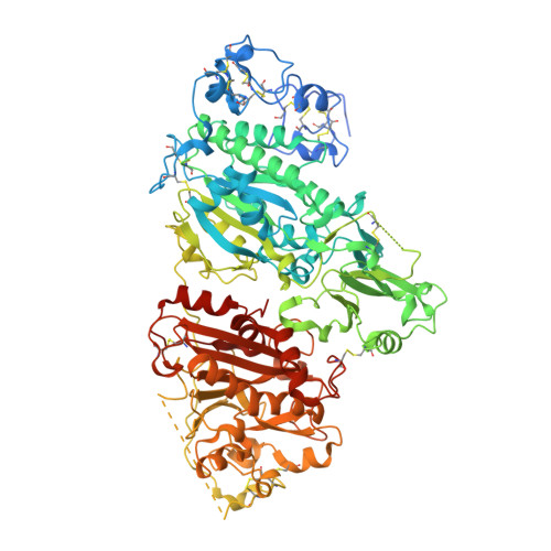
    5L0B, 5L0E, 5L0K

  • PubMed Abstract: 

    In an effort to develop a novel therapeutic agent aimed at addressing the unmet need of patients with osteoarthritis pain, we set out to develop an inhibitor for autotaxin with excellent potency and physical properties to allow for the clinical investigation of autotaxin-induced nociceptive and neuropathic pain. An initial hit identification campaign led to an aminopyrimidine series with an autotaxin IC50 of 500 nM. X-ray crystallography enabled the optimization to a lead compound that demonstrated favorable potency (IC50 = 2 nM), PK properties, and a robust PK/PD relationship.


  • Organizational Affiliation
    • Lilly Research Laboratories , A Division of Eli Lilly and Company, Indianapolis, Indiana 46285, United States.

Macromolecule Content 

  • Total Structure Weight: 204.06 kDa 
  • Atom Count: 13,448 
  • Modeled Residue Count: 1,560 
  • Deposited Residue Count: 1,742 
  • Unique protein chains: 1

Macromolecules

Find similar proteins by:|  3D Structure
Entity ID: 1
MoleculeChains  Sequence LengthOrganismDetailsImage
Ectonucleotide pyrophosphatase/phosphodiesterase family member 2
A, B
871Rattus norvegicusMutation(s): 2 
Gene Names: Enpp2AtxNpps2
EC: 3.1.4.39 (PDB Primary Data), 3.1.4.4 (UniProt)
UniProt
Find proteins for Q64610 (Rattus norvegicus)
Explore Q64610 
Go to UniProtKB:  Q64610
Entity Groups
Sequence Clusters30% Identity50% Identity70% Identity90% Identity95% Identity100% Identity
UniProt GroupQ64610
Glycosylation
Glycosylation Sites: 2Go to GlyGen: Q64610-1
Sequence Annotations
Expand
Reference Sequence

Oligosaccharides

Help  
Entity ID: 2
MoleculeChains Length2D Diagram GlycosylationD Interactions
alpha-D-mannopyranose-(1-2)-alpha-D-mannopyranose-(1-3)-[alpha-D-mannopyranose-(1-2)-alpha-D-mannopyranose-(1-6)]alpha-D-mannopyranose-(1-6)-[alpha-D-mannopyranose-(1-3)]beta-D-mannopyranose-(1-4)-2-acetamido-2-deoxy-beta-D-glucopyranose-(1-4)-2-acetamido-2-deoxy-beta-D-glucopyranose
C
9N-Glycosylation
Glycosylation Resources
GlyTouCan: G37135JQ
GlyCosmos: G37135JQ
GlyGen: G37135JQ
Entity ID: 3
MoleculeChains Length2D Diagram GlycosylationD Interactions
alpha-D-mannopyranose-(1-2)-alpha-D-mannopyranose-(1-3)-alpha-D-mannopyranose-(1-6)-beta-D-mannopyranose-(1-4)-2-acetamido-2-deoxy-beta-D-glucopyranose-(1-4)-2-acetamido-2-deoxy-beta-D-glucopyranose
D
6N-Glycosylation
Glycosylation Resources
GlyTouCan: G47410OF
GlyCosmos: G47410OF
GlyGen: G47410OF

Small Molecules

Ligands 4 Unique
IDChains Name / Formula / InChI Key2D Diagram3D Interactions
6ZM

Query on 6ZM


Download:Ideal Coordinates CCD File
F [auth A],
L [auth B]
1-{2-[(2,3-dihydro-1H-inden-2-yl)amino]-7,8-dihydropyrido[4,3-d]pyrimidin-6(5H)-yl}ethan-1-one
C18 H20 N4 O
YZUQRESBRMOGBQ-UHFFFAOYSA-N
NAG

Query on NAG


Download:Ideal Coordinates CCD File
E [auth A],
K [auth B]
2-acetamido-2-deoxy-beta-D-glucopyranose
C8 H15 N O6
OVRNDRQMDRJTHS-FMDGEEDCSA-N
ZN

Query on ZN


Download:Ideal Coordinates CCD File
G [auth A]
H [auth A]
I [auth A]
M [auth B]
N [auth B]
G [auth A],
H [auth A],
I [auth A],
M [auth B],
N [auth B],
O [auth B]
ZINC ION
Zn
PTFCDOFLOPIGGS-UHFFFAOYSA-N
CL

Query on CL


Download:Ideal Coordinates CCD File
J [auth A],
P [auth B]
CHLORIDE ION
Cl
VEXZGXHMUGYJMC-UHFFFAOYSA-M
Binding Affinity Annotations 
IDSourceBinding Affinity
6ZM BindingDB:  5L0B IC50: min: 520, max: 1070 (nM) from 2 assay(s)

Experimental Data & Validation

Experimental Data

  • Method: X-RAY DIFFRACTION
  • Resolution: 2.41 Å
  • R-Value Free:  0.209 (Depositor), 0.223 (DCC) 
  • R-Value Work:  0.176 (Depositor), 0.186 (DCC) 
  • R-Value Observed: 0.176 (Depositor) 
Space Group: C 1 2 1
Unit Cell:
Length ( Å )Angle ( ˚ )
a = 182.827α = 90
b = 96.733β = 112.86
c = 136.284γ = 90
Software Package:
Software NamePurpose
SCALAdata scaling
BUSTERrefinement
PDB_EXTRACTdata extraction
XDSdata reduction
Cootmodel building

Structure Validation

View Full Validation Report


Ligand Structure Quality Assessment 


Entry History 

Deposition Data

  • Released Date: 2016-08-10 
  • Deposition Author(s): Durbin, J.D.

Revision History  (Full details and data files)

  • Version 1.0: 2016-08-10
    Type: Initial release
  • Version 1.1: 2016-10-05
    Changes: Database references
  • Version 2.0: 2020-07-29
    Type: Remediation
    Reason: Carbohydrate remediation
    Changes: Atomic model, Data collection, Derived calculations, Structure summary
  • Version 2.1: 2024-10-30
    Changes: Data collection, Database references, Derived calculations, Structure summary