


Crystal Structure of Quorum-Sensing Transcriptional Activator from Yersinia enterocolitica
Kim, Y., Chhor, G., Jedrzejczak, R., Winans, S.C., Joachimiak, A., Center for Structural Genomics of Infectious Diseases (CSGID)To be published.
Experimental Data Snapshot
Starting Model: experimental
View more details
Entity ID: 1 | |||||
|---|---|---|---|---|---|
| Molecule | Chains | Sequence Length | Organism | Details | Image |
| Quorum-sensing transcriptional activator | 179 | Yersinia enterocolitica subsp. enterocolitica 8081 | Mutation(s): 0 Gene Names: yenR, YE1599 | 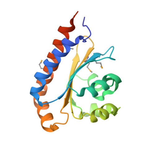 | |
UniProt | |||||
Entity Groups | |||||
| Sequence Clusters | 30% Identity50% Identity70% Identity90% Identity95% Identity100% Identity | ||||
| UniProt Group | P54295 | ||||
Sequence AnnotationsExpand | |||||
Reference Sequence | |||||
| Ligands 4 Unique | |||||
|---|---|---|---|---|---|
| ID | Chains | Name / Formula / InChI Key | 2D Diagram | 3D Interactions | |
| 482 Download:Ideal Coordinates CCD File | C [auth A], K [auth B] | 3-oxo-N-[(3S)-2-oxotetrahydrofuran-3-yl]hexanamide C10 H15 N O4 YRYOXRMDHALAFL-QMMMGPOBSA-N |  | ||
| SO4 Download:Ideal Coordinates CCD File | D [auth A], L [auth B], M [auth B] | SULFATE ION O4 S QAOWNCQODCNURD-UHFFFAOYSA-L |  | ||
| EDO Download:Ideal Coordinates CCD File | E [auth A] F [auth A] G [auth A] N [auth B] O [auth B] | 1,2-ETHANEDIOL C2 H6 O2 LYCAIKOWRPUZTN-UHFFFAOYSA-N |  | ||
| ACY Download:Ideal Coordinates CCD File | H [auth A], I [auth A], J [auth B], Q [auth B] | ACETIC ACID C2 H4 O2 QTBSBXVTEAMEQO-UHFFFAOYSA-N |  | ||
| Modified Residues 1 Unique | |||||
|---|---|---|---|---|---|
| ID | Chains | Type | Formula | 2D Diagram | Parent |
| MSE Query on MSE | A, B | L-PEPTIDE LINKING | C5 H11 N O2 Se |  | MET |
| Length ( Å ) | Angle ( ˚ ) |
|---|---|
| a = 63.878 | α = 90 |
| b = 122.775 | β = 90 |
| c = 107.603 | γ = 90 |
| Software Name | Purpose |
|---|---|
| PHENIX | refinement |
| HKL-3000 | data reduction |
| HKL-3000 | data scaling |
| HKL-3000 | phasing |
| Funding Organization | Location | Grant Number |
|---|---|---|
| National Institutes of Health/National Institute Of Allergy and Infectious Diseases (NIH/NIAID) | United States | NIAID-DMID-NIHA12011124 |


