5L05

Crystal structure of catalase-peroxidase KATG of burkholderia pseudomallei treated with INH


Experimental Data Snapshot

  • Method: X-RAY DIFFRACTION
  • Resolution: 1.70 Å
  • R-Value Free: 0.176 
  • R-Value Work: 0.145 
  • R-Value Observed: 0.148 

wwPDB Validation   3D Report Full Report


Ligand Structure Quality Assessment 


This is version 2.1 of the entry. See complete history


Literature

Catalase-peroxidase KatG of Burkholderia pseudomallei at 1.7A resolution.

Carpena, X.Loprasert, S.Mongkolsuk, S.Switala, J.Loewen, P.C.Fita, I.

(2003) J Mol Biol 327: 475-489

  • DOI: https://doi.org/10.1016/s0022-2836(03)00122-0
  • Primary Citation of Related Structures:  
    5L05

  • PubMed Abstract: 

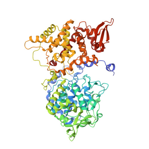
    The catalase-peroxidase encoded by katG of Burkholderia pseudomallei (BpKatG) is 65% identical with KatG of Mycobacterium tuberculosis, the enzyme responsible for the activation of isoniazid as an antibiotic. The structure of a complex of BpKatG with an unidentified ligand, has been solved and refined at 1.7A resolution using X-ray synchrotron data collected from crystals flash-cooled with liquid nitrogen. The crystallographic agreement factors R and R(free) are 15.3% and 18.6%, respectively. The crystallized enzyme is a dimer with one modified heme group and one metal ion, likely sodium, per subunit. The modification on the heme group involves the covalent addition of two or three atoms, likely a perhydroxy group, to the secondary carbon atom of the vinyl group on ring I. The added group can form hydrogen bonds with two water molecules that are also in contact with the active-site residues Trp111 and His112, suggesting that the modification may have a catalytic role. The heme modification is in close proximity to an unusual covalent adduct among the side-chains of Trp111, Tyr238 and Met264. In addition, Trp111 appears to be oxidized on C(delta1) of the indole ring. The main channel, providing access of substrate hydrogen peroxide to the heme, contains a region of unassigned electron density consistent with the binding of a pyridine nucleotide-like molecule. An interior cavity, containing the sodium ion and an additional region of unassigned density, is evident adjacent to the adduct and is accessible to the outside through a second funnel-shaped channel. A large cleft in the side of the subunit is evident and may be a potential substrate-binding site with a clear pathway for electron transfer to the active-site heme group through the adduct.


  • Organizational Affiliation

    CID-CSIC, Jordi-Girona 18-26, 08034 Barcelona, Spain.


Macromolecules
Find similar proteins by:  (by identity cutoff)  |  3D Structure
Entity ID: 1
MoleculeChains Sequence LengthOrganismDetailsImage
Catalase-peroxidase
A, B
728Burkholderia pseudomallei 1710bMutation(s): 0 
Gene Names: katGBURPS1710b_3366
EC: 1.11.1.21
UniProt
Find proteins for Q3JNW6 (Burkholderia pseudomallei (strain 1710b))
Explore Q3JNW6 
Go to UniProtKB:  Q3JNW6
Entity Groups  
Sequence Clusters30% Identity50% Identity70% Identity90% Identity95% Identity100% Identity
UniProt GroupQ3JNW6
Sequence Annotations
Expand
  • Reference Sequence
Small Molecules
Ligands 4 Unique
IDChains Name / Formula / InChI Key2D Diagram3D Interactions
HP5
Query on HP5

Download Ideal Coordinates CCD File 
C [auth A],
H [auth B]
Peroxidized Heme Form 2
C34 H32 Fe N4 O6
NXBKHEUPFTWXKQ-FPZTWHSASA-L
MPD
Query on MPD

Download Ideal Coordinates CCD File 
F [auth A],
G [auth A],
K [auth B]
(4S)-2-METHYL-2,4-PENTANEDIOL
C6 H14 O2
SVTBMSDMJJWYQN-YFKPBYRVSA-N
OXY
Query on OXY

Download Ideal Coordinates CCD File 
E [auth A],
J [auth B]
OXYGEN MOLECULE
O2
MYMOFIZGZYHOMD-UHFFFAOYSA-N
NA
Query on NA

Download Ideal Coordinates CCD File 
D [auth A],
I [auth B]
SODIUM ION
Na
FKNQFGJONOIPTF-UHFFFAOYSA-N
Experimental Data & Validation

Experimental Data

  • Method: X-RAY DIFFRACTION
  • Resolution: 1.70 Å
  • R-Value Free: 0.176 
  • R-Value Work: 0.145 
  • R-Value Observed: 0.148 
  • Space Group: P 21 21 21
Unit Cell:
Length ( Å )Angle ( ˚ )
a = 100.94α = 90
b = 115.62β = 90
c = 175.2γ = 90
Software Package:
Software NamePurpose
REFMACrefinement
DENZOdata reduction
SCALEPACKdata scaling
AMoREphasing

Structure Validation

View Full Validation Report


Ligand Structure Quality Assessment 


Entry History 

Deposition Data

  • Released Date: 2016-08-24 
  • Deposition Author(s): Loewen, P.C.
  • This entry supersedes: 1MWV

Revision History  (Full details and data files)

  • Version 1.0: 2016-08-24
    Type: Initial release
  • Version 1.1: 2017-11-22
    Changes: Data collection, Derived calculations, Refinement description, Structure summary
  • Version 2.0: 2018-03-28
    Changes: Atomic model, Data collection, Derived calculations, Refinement description
  • Version 2.1: 2023-10-04
    Changes: Data collection, Database references, Derived calculations, Refinement description