5KZ8 | pdb_00005kz8

Mark2 complex with 7-[(1S)-1-(4-fluorophenyl)ethyl]-5,5-dimethyl-2-(3-pyridylamino)pyrrolo[2,3-d]pyrimidin-6-one


Experimental Data Snapshot

  • Method: X-RAY DIFFRACTION
  • Resolution: 3.21 Å
  • R-Value Free: 
    0.290 (Depositor), 0.310 (DCC) 
  • R-Value Work: 
    0.219 (Depositor), 0.229 (DCC) 
  • R-Value Observed: 
    0.222 (Depositor) 

wwPDB Validation 3D Report Full Report

Validation slider image for 5KZ8

Ligand Structure Quality Assessment 


This is version 1.1 of the entry. See complete history

Literature

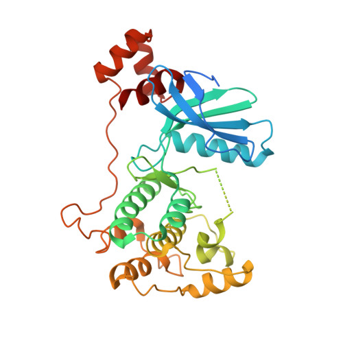
Structure guided design of a series of selective pyrrolopyrimidinone MARK inhibitors.

Katz, J.D.Haidle, A.Childers, K.K.Zabierek, A.A.Jewell, J.P.Hou, Y.Altman, M.D.Szewczak, A.Chen, D.Harsch, A.Hayashi, M.Warren, L.Hutton, M.Nuthall, H.Su, H.P.Munshi, S.Stanton, M.G.Davies, I.W.Munoz, B.Northrup, A.

(2017) Bioorg Med Chem Lett 27: 114-120

  • DOI: https://doi.org/10.1016/j.bmcl.2016.08.068
  • Primary Citation Related Structures: 
    5KZ7, 5KZ8

  • PubMed Abstract: 

    The initial structure activity relationships around an isoindoline uHTS hit will be described. Information gleaned from ligand co-crystal structures allowed for rapid refinements in both MARK potency and kinase selectivity. These efforts allowed for the identification of a compound with properties suitable for use as an in vitro tool compound for validation studies on MARK as a viable target for Alzheimer's disease.


  • Organizational Affiliation
    • Department of Chemistry, Merck & Co., Inc., 33 Avenue Louis Pasteur, Boston, MA 02115, USA. Electronic address: jason_katz2@merck.com.

Macromolecule Content 

  • Total Structure Weight: 80.88 kDa 
  • Atom Count: 4,888 
  • Modeled Residue Count: 602 
  • Deposited Residue Count: 692 
  • Unique protein chains: 1

Macromolecules

Find similar proteins by:|  3D Structure
Entity ID: 1
MoleculeChains  Sequence LengthOrganismDetailsImage
Serine/threonine-protein kinase MARK2
A, B
346Homo sapiensMutation(s): 0 
Gene Names: MARK2EMK1
EC: 2.7.11.1 (PDB Primary Data), 2.7.11.26 (PDB Primary Data)
UniProt & NIH Common Fund Data Resources
Find proteins for Q7KZI7 (Homo sapiens)
Explore Q7KZI7 
Go to UniProtKB:  Q7KZI7
PHAROS:  Q7KZI7
GTEx:  ENSG00000072518 
Entity Groups
Sequence Clusters30% Identity50% Identity70% Identity90% Identity95% Identity100% Identity
UniProt GroupQ7KZI7
Sequence Annotations
Expand
Reference Sequence

Small Molecules

Ligands 1 Unique
IDChains Name / Formula / InChI Key2D Diagram3D Interactions
6Z5

Query on 6Z5


Download:Ideal Coordinates CCD File
C [auth A],
D [auth B]
5,5-dimethyl-7-[(1~{S})-4-oxidanyl-1~{H}-inden-1-yl]-2-phenylazanyl-pyrrolo[2,3-d]pyrimidin-6-one
C23 H20 N4 O2
XYYZSCWSVMFLOM-SFHVURJKSA-N

Experimental Data & Validation

Experimental Data

  • Method: X-RAY DIFFRACTION
  • Resolution: 3.21 Å
  • R-Value Free:  0.290 (Depositor), 0.310 (DCC) 
  • R-Value Work:  0.219 (Depositor), 0.229 (DCC) 
  • R-Value Observed: 0.222 (Depositor) 
Space Group: P 61
Unit Cell:
Length ( Å )Angle ( ˚ )
a = 120.416α = 90
b = 120.416β = 90
c = 99.457γ = 120
Software Package:
Software NamePurpose
HKL-2000data collection
HKL-2000data scaling
BUSTER-TNTrefinement
PDB_EXTRACTdata extraction
HKL-2000data reduction
BUSTERphasing

Structure Validation

View Full Validation Report


Ligand Structure Quality Assessment 


Entry History 

Deposition Data

Revision History  (Full details and data files)

  • Version 1.0: 2017-05-31
    Type: Initial release
  • Version 1.1: 2024-03-06
    Changes: Data collection, Database references