5KUB | pdb_00005kub

Bacillus cereus DNA glycosylase AlkD bound to 7-methylguanine nucleobase and DNA containing an oxocarbenium-intermediate analog


Experimental Data Snapshot

  • Method: X-RAY DIFFRACTION
  • Resolution: 1.73 Å
  • R-Value Free: 
    0.180 (Depositor), 0.182 (DCC) 
  • R-Value Work: 
    0.141 (Depositor), 0.143 (DCC) 
  • R-Value Observed: 
    0.143 (Depositor) 

Starting Model: experimental
View more details

wwPDB Validation 3D Report Full Report

Validation slider image for 5KUB

Ligand Structure Quality Assessment 


This is version 1.3 of the entry. See complete history

Literature

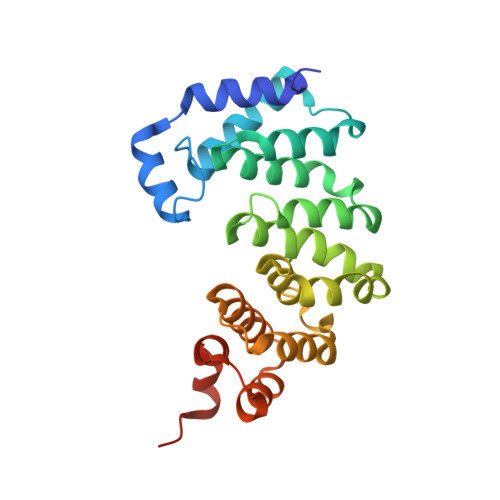
A Catalytic Role for C-H/ pi Interactions in Base Excision Repair by Bacillus cereus DNA Glycosylase AlkD.

Parsons, Z.D.Bland, J.M.Mullins, E.A.Eichman, B.F.

(2016) J Am Chem Soc 138: 11485-11488

  • DOI: https://doi.org/10.1021/jacs.6b07399
  • Primary Citation Related Structures: 
    5KUB

  • PubMed Abstract: 

    DNA glycosylases protect genomic integrity by locating and excising aberrant nucleobases. Substrate recognition and excision usually take place in an extrahelical conformation, which is often stabilized by π-stacking interactions between the lesion nucleobase and aromatic side chains in the glycosylase active site. Bacillus cereus AlkD is the only DNA glycosylase known to catalyze base excision without extruding the damaged nucleotide from the DNA helix. Instead of contacting the nucleobase itself, the AlkD active site interacts with the lesion deoxyribose through a series of C-H/π interactions. These interactions are ubiquitous in protein structures, but evidence for their catalytic significance in enzymology is lacking. Here, we show that the C-H/π interactions between AlkD and the lesion deoxyribose participate in catalysis of glycosidic bond cleavage. This is the first demonstration of a catalytic role for C-H/π interactions as intermolecular forces important to DNA repair.


  • Organizational Affiliation
    • Department of Biological Sciences and Center for Structural Biology, Vanderbilt University , Nashville, Tennessee 37232, United States.

Macromolecule Content 

  • Total Structure Weight: 35.92 kDa 
  • Atom Count: 2,781 
  • Modeled Residue Count: 255 
  • Deposited Residue Count: 265 
  • Unique protein chains: 1
  • Unique nucleic acid chains: 2

Macromolecules


Find similar proteins by:|  3D Structure
Entity ID: 1
MoleculeChains  Sequence LengthOrganismDetailsImage
DNA-7-methylguanine glycosylase241Bacillus cereusMutation(s): 0 
Gene Names: bcere0015_46090
EC: 3.2.2
UniProt
Find proteins for Q816E8 (Bacillus cereus (strain ATCC 14579 / DSM 31 / CCUG 7414 / JCM 2152 / NBRC 15305 / NCIMB 9373 / NCTC 2599 / NRRL B-3711))
Explore Q816E8 
Go to UniProtKB:  Q816E8
Entity Groups
Sequence Clusters30% Identity50% Identity70% Identity90% Identity95% Identity100% Identity
UniProt GroupQ816E8
Sequence Annotations
Expand
Reference Sequence
Find similar nucleic acids by:  Sequence
Entity ID: 2
MoleculeChains LengthOrganismImage
DNA (5'-D(*CP*CP*CP*GP*AP*(NRI)P*AP*GP*TP*CP*CP*G)-3')12synthetic construct
Sequence Annotations
Expand
Reference Sequence
Find similar nucleic acids by:  Sequence
Entity ID: 3
MoleculeChains LengthOrganismImage
DNA (5'-D(*CP*GP*GP*AP*CP*TP*CP*TP*CP*GP*GP*G)-3')12synthetic construct
Sequence Annotations
Expand
Reference Sequence

Small Molecules

Ligands 1 Unique
IDChains Name / Formula / InChI Key2D Diagram3D Interactions
MY6

Query on MY6


Download:Ideal Coordinates CCD File
D [auth B]2-amino-7-methyl-1,7-dihydro-6H-purin-6-one
C6 H7 N5 O
FZWGECJQACGGTI-UHFFFAOYSA-N

Experimental Data & Validation

Experimental Data

  • Method: X-RAY DIFFRACTION
  • Resolution: 1.73 Å
  • R-Value Free:  0.180 (Depositor), 0.182 (DCC) 
  • R-Value Work:  0.141 (Depositor), 0.143 (DCC) 
  • R-Value Observed: 0.143 (Depositor) 
Space Group: P 1 21 1
Unit Cell:
Length ( Å )Angle ( ˚ )
a = 38.45α = 90
b = 93.603β = 112.54
c = 47.991γ = 90
Software Package:
Software NamePurpose
HKL-2000data scaling
PHENIXrefinement
PDB_EXTRACTdata extraction
HKL-2000data reduction
PHENIXphasing

Structure Validation

View Full Validation Report


Ligand Structure Quality Assessment 


Entry History 

& Funding Information

Deposition Data


Funding OrganizationLocationGrant Number
National Science Foundation (NSF, United States)United StatesMCB-1122098
National Science Foundation (NSF, United States)United StatesMCB-1517695
National Institutes of Health/National Institute of Environmental Health Sciences (NIH/NIEHS)United StatesR01 ES019625

Revision History  (Full details and data files)

  • Version 1.0: 2016-09-14
    Type: Initial release
  • Version 1.1: 2016-09-28
    Changes: Database references
  • Version 1.2: 2019-11-27
    Changes: Author supporting evidence, Derived calculations
  • Version 1.3: 2023-10-04
    Changes: Data collection, Database references, Refinement description