5KRT | pdb_00005krt

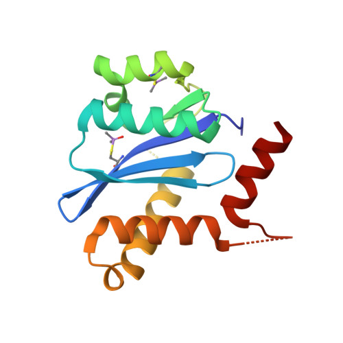
HIV-1 Integrase Catalytic Core Domain (CCD) in Complex with a Fragment-Derived Allosteric Inhibitor


Experimental Data Snapshot

  • Method: X-RAY DIFFRACTION
  • Resolution: 1.65 Å
  • R-Value Free: 
    0.195 (Depositor), 0.199 (DCC) 
  • R-Value Work: 
    0.185 (Depositor), 0.186 (DCC) 
  • R-Value Observed: 
    0.186 (Depositor) 

wwPDB Validation 3D Report Full Report

Validation slider image for 5KRT

Ligand Structure Quality Assessment 


This is version 1.5 of the entry. See complete history. 

Literature

A New Class of Allosteric HIV-1 Integrase Inhibitors Identified by Crystallographic Fragment Screening of the Catalytic Core Domain.

Patel, D., Antwi, J., Koneru, P.C., Serrao, E., Forli, S., Kessl, J.J., Feng, L., Deng, N., Levy, R.M., Fuchs, J.R., Olson, A.J., Engelman, A.N., Bauman, J.D., Kvaratskhelia, M., Arnold, E.

(2016) J Biol Chem 291: 23569-23577

  • DOI: https://doi.org/10.1074/jbc.M116.753384
  • Primary Citation Related Structures: 
    5KRS, 5KRT

  • PubMed Abstract: 

    HIV-1 integrase (IN) is essential for virus replication and represents an important multifunctional therapeutic target. Recently discovered quinoline-based allosteric IN inhibitors (ALLINIs) potently impair HIV-1 replication and are currently in clinical trials. ALLINIs exhibit a multimodal mechanism of action by inducing aberrant IN multimerization during virion morphogenesis and by competing with IN for binding to its cognate cellular cofactor LEDGF/p75 during early steps of HIV-1 infection. However, quinoline-based ALLINIs impose a low genetic barrier for the evolution of resistant phenotypes, which highlights a need for discovery of second-generation inhibitors. Using crystallographic screening of a library of 971 fragments against the HIV-1 IN catalytic core domain (CCD) followed by a fragment expansion approach, we have identified thiophenecarboxylic acid derivatives that bind at the CCD-CCD dimer interface at the principal lens epithelium-derived growth factor (LEDGF)/p75 binding pocket. The most active derivative (5) inhibited LEDGF/p75-dependent HIV-1 IN activity in vitro with an IC 50 of 72 μm and impaired HIV-1 infection of T cells at an EC 50 of 36 μm The identified lead compound, with a relatively small molecular weight (221 Da), provides an optimal building block for developing a new class of inhibitors. Furthermore, although structurally distinct thiophenecarboxylic acid derivatives target a similar pocket at the IN dimer interface as the quinoline-based ALLINIs, the lead compound, 5, inhibited IN mutants that confer resistance to quinoline-based compounds. Collectively, our findings provide a plausible path for structure-based development of second-generation ALLINIs.


  • Organizational Affiliation: 
    • From the Center for Advanced Biotechnology and Medicine, Rutgers University, Piscataway, New Jersey 08854.

Macromolecule Content 

  • Total Structure Weight: 17.51 kDa 
  • Atom Count: 1,282 
  • Modeled Residue Count: 145 
  • Deposited Residue Count: 153 
  • Unique protein chains: 1

Macromolecules

Find similar proteins by:|  3D Structure
Entity ID: 1
MoleculeChains  Sequence LengthOrganismDetailsImage
Integrase153Human immunodeficiency virus 1Mutation(s): 1 
UniProt
Find proteins for P12497 (Human immunodeficiency virus type 1 group M subtype B (isolate NY5))
Explore P12497 
Go to UniProtKB:  P12497
Entity Groups
Sequence Clusters30% Identity50% Identity70% Identity90% Identity95% Identity100% Identity
UniProt GroupP12497
Sequence Annotations
Expand
Reference Sequence

Small Molecules

Ligands 1 Unique
IDChains Name / Formula / InChI Key2D Diagram3D Interactions
6W6

Query on 6W6


Download:Ideal Coordinates CCD File
B [auth A],
C [auth A]
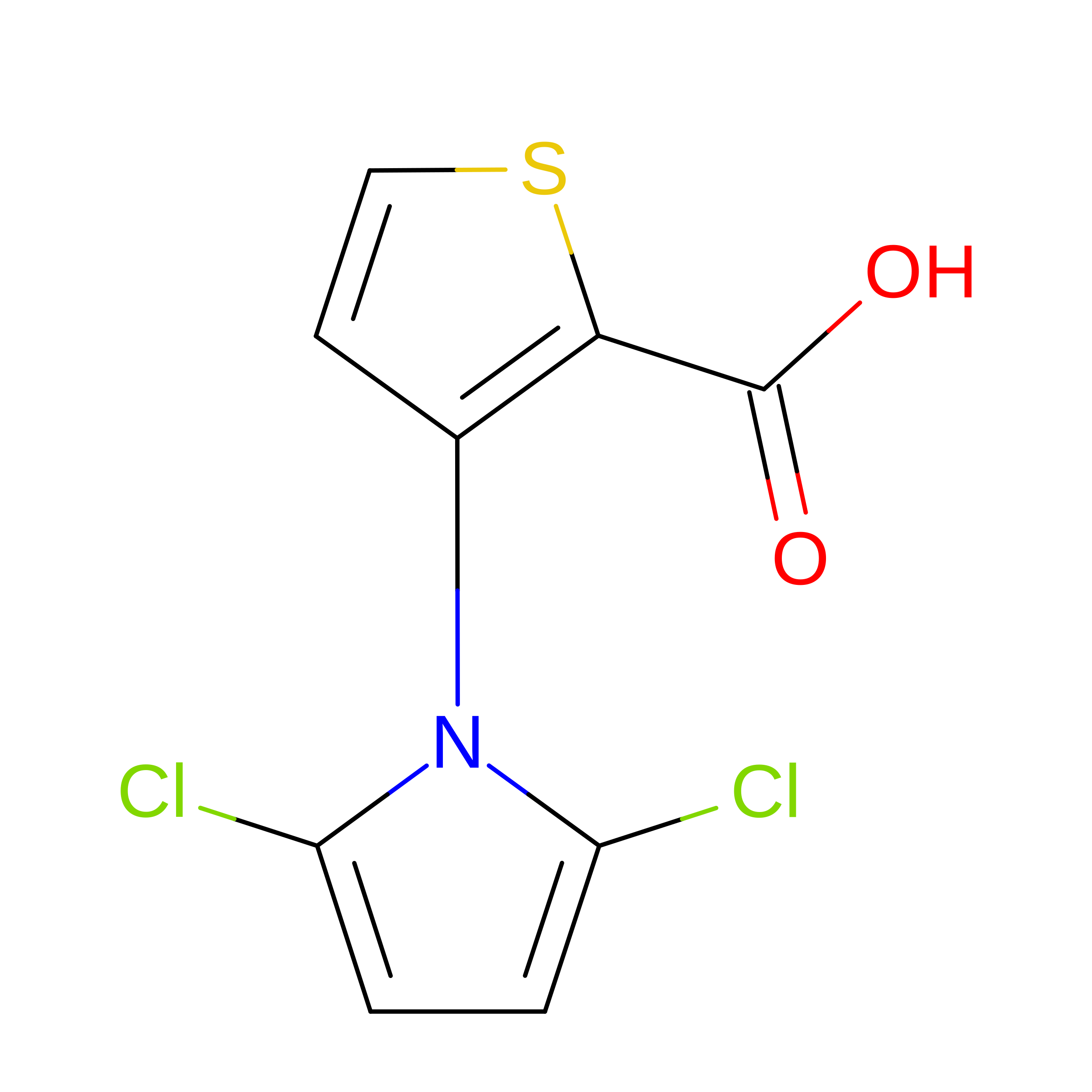
3-[2,5-bis(chloranyl)pyrrol-1-yl]thiophene-2-carboxylic acid
C9 H5 Cl2 N O2 S
BQBZZKAKSYADDO-UHFFFAOYSA-N
Non-standard Monomers  1 Unique
IDChains TypeFormula2D Diagram
CAF
Query on CAF
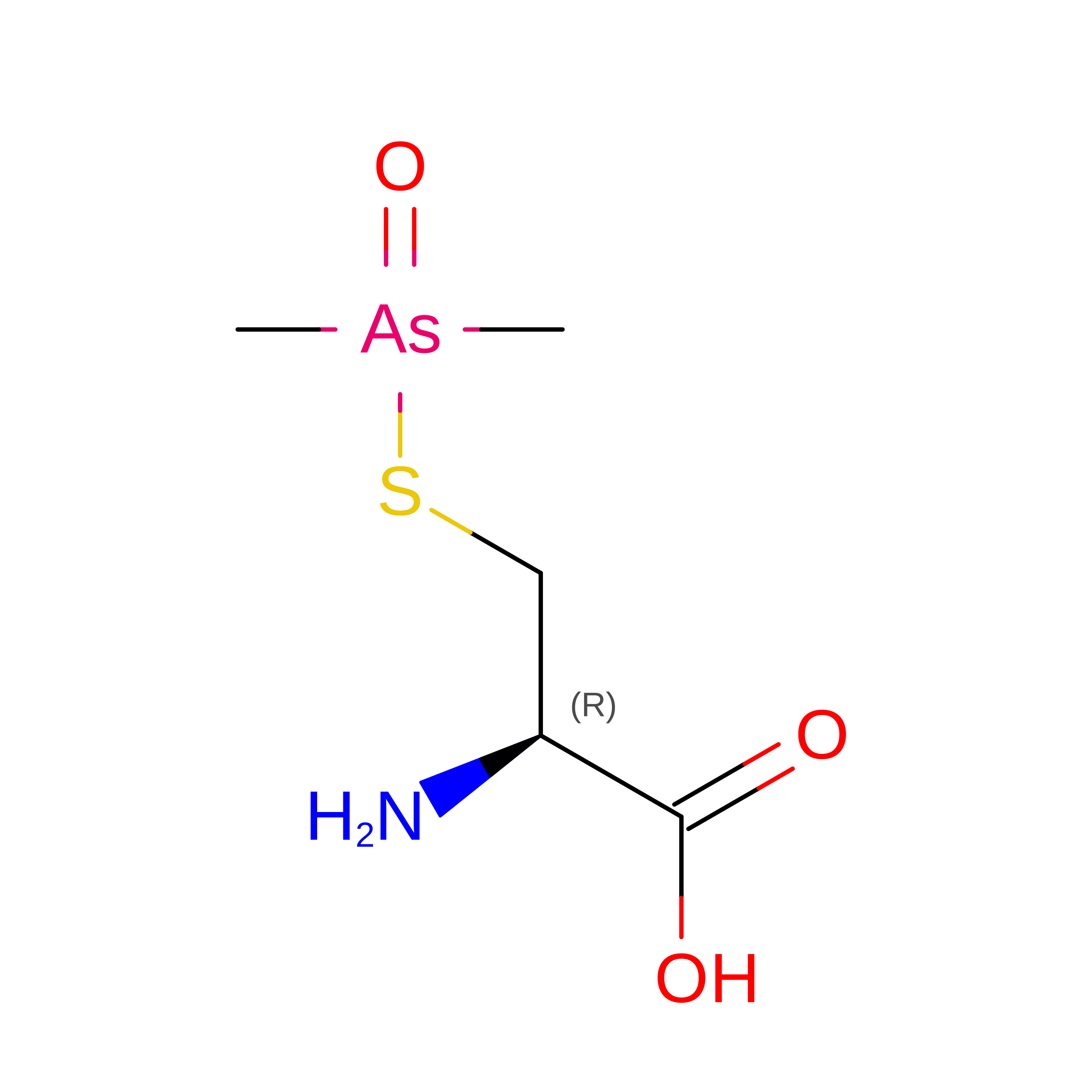
L-PEPTIDE LINKINGC5 H12 As N O3 S

Experimental Data & Validation

Experimental Data

  • Method: X-RAY DIFFRACTION
  • Resolution: 1.65 Å
  • R-Value Free:  0.195 (Depositor), 0.199 (DCC) 
  • R-Value Work:  0.185 (Depositor), 0.186 (DCC) 
  • R-Value Observed: 0.186 (Depositor) 
Space Group: P 31 2 1
Unit Cell:
Length ( Å )Angle ( ˚ )
a = 72.069α = 90
b = 72.069β = 90
c = 66.348γ = 120
Software Package:
Software NamePurpose
PHENIXrefinement
HKL-2000data reduction
SCALEPACKdata scaling

Structure Validation

View Full Validation Report


Ligand Structure Quality Assessment 


Entry History 

& Funding Information

Deposition Data


Funding OrganizationLocationGrant Number
National Institutes of Health/National Institute of General Medical Sciences (NIH/NIGMS)United StatesGM103368, AI110310, AI110270

Revision History  (Full details and data files)

  • Version 1.0: 2016-09-28
    Type: Initial release
  • Version 1.1: 2016-10-05
    Changes: Database references
  • Version 1.2: 2016-11-16
    Changes: Database references
  • Version 1.3: 2017-09-20
    Changes: Author supporting evidence, Database references, Derived calculations
  • Version 1.4: 2019-12-25
    Changes: Author supporting evidence
  • Version 1.5: 2024-10-30
    Changes: Data collection, Database references, Structure summary