5KM8

Human Histidine Triad Nucleotide Binding Protein 2 (hHint2) Cidofovir complex

  • Classification: HYDROLASE
  • Organism(s): Homo sapiens
  • Expression System: Escherichia coli
  • Mutation(s): No 

  • Deposited: 2016-06-26 Released: 2017-06-28 
  • Deposition Author(s): Maize, K.M., Finzel, B.C.
  • Funding Organization(s): National Institutes of Health/National Institute of General Medical Sciences (NIH/NIGMS)

Experimental Data Snapshot

  • Method: X-RAY DIFFRACTION
  • Resolution: 2.00 Å
  • R-Value Free: 0.212 
  • R-Value Work: 0.161 
  • R-Value Observed: 0.163 

wwPDB Validation   3D Report Full Report


Ligand Structure Quality Assessment 


This is version 1.4 of the entry. See complete history


Literature

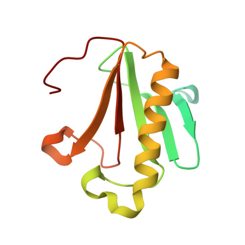
A Crystal Structure Based Guide to the Design of Human Histidine Triad Nucleotide Binding Protein 1 (hHint1) Activated ProTides.

Maize, K.M.Shah, R.Strom, A.Kumarapperuma, S.Zhou, A.Wagner, C.R.Finzel, B.C.

(2017) Mol Pharm 14: 3987-3997

  • DOI: 10.1021/acs.molpharmaceut.7b00664
  • Primary Citation of Related Structures:  
    5KLY, 5KLZ, 5KM0, 5KM1, 5KM2, 5KM3, 5KM4, 5KM5, 5KM6, 5KM8, 5KM9, 5KMA, 5KMB, 5WA8, 5WA9, 6B42

  • PubMed Abstract: 
  • Nucleotide analogues that incorporate a metabolically labile nucleoside phosphoramidate (a ProTide) have found utility as prodrugs. In humans, ProTides can be cleaved by human histidine triad nucleotide binding protein 1 (hHint1) to expose the nucleotide monophosphate ...

    Nucleotide analogues that incorporate a metabolically labile nucleoside phosphoramidate (a ProTide) have found utility as prodrugs. In humans, ProTides can be cleaved by human histidine triad nucleotide binding protein 1 (hHint1) to expose the nucleotide monophosphate. Activation by this route circumvents highly selective nucleoside kinases that limit the use of nucleosides as prodrugs. To better understand the diversity of potential substrates of hHint1, we created and studied a series of phosphoramidate nucleosides. Using a combination of enzyme kinetics, X-ray crystallography, and isothermal titration calorimetry with both wild-type and inactive mutant enzymes, we have been able to explore the energetics of substrate binding and establish a structural basis for catalytic efficiency. Diverse nucleobases are well tolerated, but portions of the ribose are needed to position substrates for catalysis. Beneficial characteristics of the amine leaving group are also revealed. Structural principles revealed by these results may be exploited to tune the rate of substrate hydrolysis to strategically alter the intracellular release of the product nucleoside monophosphate from the ProTide.


    Organizational Affiliation

    Department of Medicinal Chemistry, University of Minnesota , Minneapolis, Minnesota 55455, United States.


Macromolecules
Find similar proteins by:  (by identity cutoff)  |  3D Structure
Entity ID: 1
MoleculeChainsSequence LengthOrganismDetailsImage
Histidine triad nucleotide-binding protein 2, mitochondrialA, B142Homo sapiensMutation(s): 0 
Gene Names: HINT2
EC: 3 (PDB Primary Data), 3.9.1 (UniProt)
UniProt & NIH Common Fund Data Resources
Find proteins for Q9BX68 (Homo sapiens)
Explore Q9BX68 
Go to UniProtKB:  Q9BX68
PHAROS:  Q9BX68
Entity Groups  
Sequence Clusters30% Identity50% Identity70% Identity90% Identity95% Identity100% Identity
UniProt GroupQ9BX68
Protein Feature View
Expand
  • Reference Sequence
Small Molecules
Ligands 5 Unique
IDChainsName / Formula / InChI Key2D Diagram3D Interactions
L8P
Query on L8P

Download Ideal Coordinates CCD File 
E [auth B]({[(2S)-1-(4-amino-2-oxopyrimidin-1(2H)-yl)-3-hydroxypropan-2-yl]oxy}methyl)phosphonic acid
C8 H14 N3 O6 P
VWFCHDSQECPREK-LURJTMIESA-N
 Ligand Interaction
GOL
Query on GOL

Download Ideal Coordinates CCD File 
H [auth B]GLYCEROL
C3 H8 O3
PEDCQBHIVMGVHV-UHFFFAOYSA-N
 Ligand Interaction
DMS
Query on DMS

Download Ideal Coordinates CCD File 
D [auth A]DIMETHYL SULFOXIDE
C2 H6 O S
IAZDPXIOMUYVGZ-UHFFFAOYSA-N
 Ligand Interaction
EDO
Query on EDO

Download Ideal Coordinates CCD File 
F [auth B],
G [auth B]
1,2-ETHANEDIOL
C2 H6 O2
LYCAIKOWRPUZTN-UHFFFAOYSA-N
 Ligand Interaction
CL
Query on CL

Download Ideal Coordinates CCD File 
C [auth A]CHLORIDE ION
Cl
VEXZGXHMUGYJMC-UHFFFAOYSA-M
 Ligand Interaction
Experimental Data & Validation

Experimental Data

  • Method: X-RAY DIFFRACTION
  • Resolution: 2.00 Å
  • R-Value Free: 0.212 
  • R-Value Work: 0.161 
  • R-Value Observed: 0.163 
  • Space Group: P 21 21 21
Unit Cell:
Length ( Å )Angle ( ˚ )
a = 37.99α = 90
b = 73.852β = 90
c = 77.464γ = 90
Software Package:
Software NamePurpose
PHENIXrefinement
PDB_EXTRACTdata extraction
PHASERphasing
SCALAdata scaling
XDSdata reduction

Structure Validation

View Full Validation Report


Ligand Structure Quality Assessment 


Entry History & Funding Information

Deposition Data


Funding OrganizationLocationGrant Number
National Institutes of Health/National Institute of General Medical Sciences (NIH/NIGMS)United StatesGM08700

Revision History  (Full details and data files)

  • Version 1.0: 2017-06-28
    Type: Initial release
  • Version 1.1: 2017-07-26
    Changes: Author supporting evidence, Database references
  • Version 1.2: 2017-10-18
    Changes: Database references
  • Version 1.3: 2017-11-15
    Changes: Database references
  • Version 1.4: 2019-12-25
    Changes: Author supporting evidence, Database references