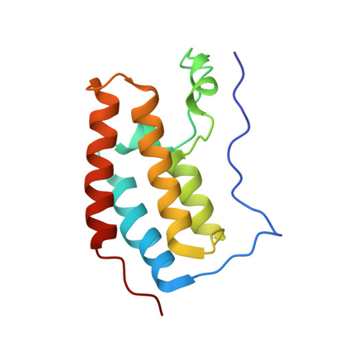
5KJ0

CRYSTAL STRUCTURE OF THE FIRST BROMODOMAIN OF HUMAN BRD4 IN COMPLEX WITH DB-1-264-2


Experimental Data Snapshot

  • Method: X-RAY DIFFRACTION
  • Resolution: 1.51 Å
  • R-Value Free: 0.170 
  • R-Value Work: 0.139 

wwPDB Validation   3D Report Full Report


Ligand Structure Quality Assessment 


This is version 1.1 of the entry. See complete history


Literature

Assessment of Bromodomain Target Engagement by a Series of BI2536 Analogues with Miniaturized BET-BRET.

Koblan, L.W.Buckley, D.L.Ott, C.J.Fitzgerald, M.E.Ember, S.W.Zhu, J.Y.Liu, S.Roberts, J.M.Remillard, D.Vittori, S.Zhang, W.Schonbrunn, E.Bradner, J.E.

(2016) ChemMedChem 11: 2575-2581

  • DOI: https://doi.org/10.1002/cmdc.201600502
  • Primary Citation of Related Structures:  
    5KJ0

  • PubMed Abstract: 

    Evaluating the engagement of a small molecule ligand with a protein target in cells provides useful information for chemical probe optimization and pharmaceutical development. While several techniques exist that can be performed in a low-throughput manner, systematic evaluation of large compound libraries remains a challenge. In-cell engagement measurements are especially useful when evaluating compound classes suspected to target multiple cellular factors. In this study we used a bioluminescent resonant energy transfer assay to assess bromodomain engagement by a compound series containing bromodomain- and kinase-biasing polypharmacophores based on the known dual BRD4 bromodomain/PLK1 kinase inhibitor BI2536. With this assay, we discovered several novel agents with bromodomain-selective specificity profiles and cellular activity. Thus, this platform aids in distinguishing molecules whose cellular activity is difficult to assess due to polypharmacologic effects.


  • Organizational Affiliation

    Center for the Science of Therapeutics, Broad Institute, Cambridge, MA, 02142, USA.


Macromolecules
Find similar proteins by:  (by identity cutoff)  |  3D Structure
Entity ID: 1
MoleculeChains Sequence LengthOrganismDetailsImage
Bromodomain-containing protein 4127Homo sapiensMutation(s): 0 
Gene Names: BRD4HUNK1
UniProt & NIH Common Fund Data Resources
Find proteins for O60885 (Homo sapiens)
Explore O60885 
Go to UniProtKB:  O60885
PHAROS:  O60885
GTEx:  ENSG00000141867 
Entity Groups  
Sequence Clusters30% Identity50% Identity70% Identity90% Identity95% Identity100% Identity
UniProt GroupO60885
Sequence Annotations
Expand
  • Reference Sequence
Small Molecules
Ligands 2 Unique
IDChains Name / Formula / InChI Key2D Diagram3D Interactions
6TB
Query on 6TB

Download Ideal Coordinates CCD File 
B [auth A]4-[[(7~{R})-8-cyclopentyl-7-ethyl-5-methyl-6-oxidanylidene-7~{H}-pteridin-2-yl]-methyl-amino]-3-methoxy-~{N}-(1-methylpiperidin-4-yl)benzamide
C29 H41 N7 O3
ZDNOQFYOJHNBGG-JOCHJYFZSA-N
EDO
Query on EDO

Download Ideal Coordinates CCD File 
C [auth A]
D [auth A]
E [auth A]
F [auth A]
G [auth A]
C [auth A],
D [auth A],
E [auth A],
F [auth A],
G [auth A],
H [auth A],
I [auth A]
1,2-ETHANEDIOL
C2 H6 O2
LYCAIKOWRPUZTN-UHFFFAOYSA-N
Binding Affinity Annotations 
IDSourceBinding Affinity
6TB Binding MOAD:  5KJ0 IC50: 130 (nM) from 1 assay(s)
Experimental Data & Validation

Experimental Data

  • Method: X-RAY DIFFRACTION
  • Resolution: 1.51 Å
  • R-Value Free: 0.170 
  • R-Value Work: 0.139 
  • Space Group: P 21 21 21
Unit Cell:
Length ( Å )Angle ( ˚ )
a = 37.11α = 90
b = 44.7β = 90
c = 77.72γ = 90
Software Package:
Software NamePurpose
XSCALEdata scaling
PHASERphasing
PHENIXrefinement
PDB_EXTRACTdata extraction
XDSdata reduction

Structure Validation

View Full Validation Report


Ligand Structure Quality Assessment 


Entry History 

Deposition Data

Revision History  (Full details and data files)

  • Version 1.0: 2017-08-09
    Type: Initial release
  • Version 1.1: 2023-09-27
    Changes: Data collection, Database references, Refinement description