5KIQ

SrpA with sialyl LewisX


Experimental Data Snapshot

  • Method: X-RAY DIFFRACTION
  • Resolution: 1.64 Å
  • R-Value Free: 0.218 
  • R-Value Work: 0.193 
  • R-Value Observed: 0.194 

wwPDB Validation   3D Report Full Report


This is version 2.1 of the entry. See complete history


Literature

Structures of the Streptococcus sanguinis SrpA Binding Region with Human Sialoglycans Suggest Features of the Physiological Ligand.

Loukachevitch, L.V.Bensing, B.A.Yu, H.Zeng, J.Chen, X.Sullam, P.M.Iverson, T.M.

(2016) Biochemistry 

  • DOI: https://doi.org/10.1021/acs.biochem.6b00704
  • Primary Citation of Related Structures:  
    5IIY, 5IJ1, 5IJ2, 5IJ3, 5KIQ

  • PubMed Abstract: 

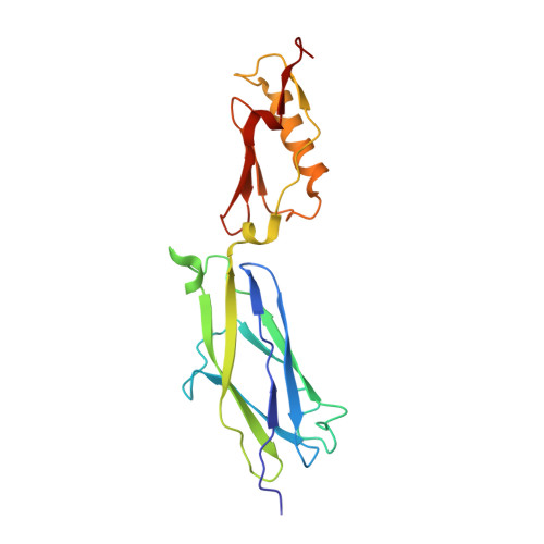
    Streptococcus sanguinis is a leading cause of bacterial infective endocarditis, a life-threatening infection of heart valves. S. sanguinis binds to human platelets with high avidity, and this adherence is likely to enhance virulence. Previous studies suggest that a serine-rich repeat adhesin termed SrpA mediates the binding of S. sanguinis to human platelets via its interaction with sialoglycans on the receptor GPIbα. However, in vitro binding assays with SrpA and defined sialoglycans failed to identify specific high-affinity ligands. To improve our understanding of the interaction between SrpA and human platelets, we determined cocrystal structures of the SrpA sialoglycan binding region (SrpA BR ) with five low-affinity ligands: three sialylated trisaccharides (sialyl-T antigen, 3'-sialyllactose, and 3'-sialyl-N-acetyllactosamine), a sialylated tetrasaccharide (sialyl-Lewis X ), and a sialyl galactose disaccharide component common to these sialoglyans. We then combined structural analysis with mutagenesis to further determine whether our observed interactions between SrpA BR and glycans are important for binding to platelets and to better map the binding site for the physiological receptor. We found that the sialoglycan binding site of SrpA BR is significantly larger than the sialoglycans cocrystallized in this study, which suggests that binding of SrpA to platelets either is multivalent or occurs via a larger, disialylated glycan.


  • Organizational Affiliation

    Division of Infectious Diseases, Veterans Affairs Medical Center, University of California, San Francisco and Northern California Institute for Research and Education , San Francisco, California 94121, United States.


Macromolecules
Find similar proteins by:  (by identity cutoff)  |  3D Structure
Entity ID: 1
MoleculeChains Sequence LengthOrganismDetailsImage
Platelet-binding glycoprotein
A, B
197Streptococcus sanguinis SK36Mutation(s): 0 
Gene Names: srpASSA_0829
UniProt
Find proteins for A3CM52 (Streptococcus sanguinis (strain SK36))
Explore A3CM52 
Go to UniProtKB:  A3CM52
Entity Groups  
Sequence Clusters30% Identity50% Identity70% Identity90% Identity95% Identity100% Identity
UniProt GroupA3CM52
Sequence Annotations
Expand
  • Reference Sequence
Oligosaccharides

Help

Entity ID: 2
MoleculeChains Length2D Diagram Glycosylation3D Interactions
N-acetyl-alpha-neuraminic acid-(2-3)-beta-D-galactopyranose-(1-4)-[alpha-L-fucopyranose-(1-3)]2-acetamido-2-deoxy-beta-D-glucopyranose
C
4N/A
Glycosylation Resources
GlyTouCan:  G00054MO
GlyCosmos:  G00054MO
GlyGen:  G00054MO
Small Molecules
Ligands 2 Unique
IDChains Name / Formula / InChI Key2D Diagram3D Interactions
ACT
Query on ACT

Download Ideal Coordinates CCD File 
G [auth A]
H [auth A]
I [auth A]
N [auth B]
O [auth B]
G [auth A],
H [auth A],
I [auth A],
N [auth B],
O [auth B],
P [auth B]
ACETATE ION
C2 H3 O2
QTBSBXVTEAMEQO-UHFFFAOYSA-M
CA
Query on CA

Download Ideal Coordinates CCD File 
D [auth A]
E [auth A]
F [auth A]
J [auth B]
K [auth B]
D [auth A],
E [auth A],
F [auth A],
J [auth B],
K [auth B],
L [auth B],
M [auth B]
CALCIUM ION
Ca
BHPQYMZQTOCNFJ-UHFFFAOYSA-N
Biologically Interesting Molecules (External Reference) 1 Unique
Experimental Data & Validation

Experimental Data

  • Method: X-RAY DIFFRACTION
  • Resolution: 1.64 Å
  • R-Value Free: 0.218 
  • R-Value Work: 0.193 
  • R-Value Observed: 0.194 
  • Space Group: C 1 2 1
Unit Cell:
Length ( Å )Angle ( ˚ )
a = 173.911α = 90
b = 46.766β = 102.73
c = 65.169γ = 90
Software Package:
Software NamePurpose
SCALEPACKdata scaling
PHENIXrefinement
PDB_EXTRACTdata extraction

Structure Validation

View Full Validation Report


Entry History & Funding Information

Deposition Data


Funding OrganizationLocationGrant Number
National Institutes of Health/National Institute Of Allergy and Infectious Diseases (NIH/NIAID)United StatesAI106987
National Institutes of Health/National Institute Of Allergy and Infectious Diseases (NIH/NIAID)United StatesAI41513
American Heart AssociationUnited States14GRNT20390021

Revision History  (Full details and data files)

  • Version 1.0: 2017-05-24
    Type: Initial release
  • Version 1.1: 2017-09-20
    Changes: Author supporting evidence
  • Version 1.2: 2019-12-11
    Changes: Author supporting evidence, Data collection
  • Version 2.0: 2020-07-29
    Type: Remediation
    Reason: Carbohydrate remediation
    Changes: Atomic model, Data collection, Derived calculations, Structure summary
  • Version 2.1: 2024-03-06
    Changes: Data collection, Database references, Structure summary