5KF6

Structure of proline utilization A from Sinorhizobium meliloti complexed with L-tetrahydrofuroic acid and NAD+ in space group P21

  • Classification: OXIDOREDUCTASE
  • Organism(s): Sinorhizobium meliloti SM11
  • Expression System: Escherichia coli
  • Mutation(s): No 

  • Deposited: 2016-06-12 Released: 2016-10-05 
  • Deposition Author(s): Tanner, J.J.
  • Funding Organization(s): National Institutes of Health/National Institute of General Medical Sciences (NIH/NIGMS), National Science Foundation (NSF, United States)

Experimental Data Snapshot

  • Method: X-RAY DIFFRACTION
  • Resolution: 1.70 Å
  • R-Value Free: 0.222 
  • R-Value Work: 0.190 
  • R-Value Observed: 0.192 

wwPDB Validation   3D Report Full Report


Ligand Structure Quality Assessment 


This is version 1.6 of the entry. See complete history


Literature

Structures of Proline Utilization A (PutA) Reveal the Fold and Functions of the Aldehyde Dehydrogenase Superfamily Domain of Unknown Function.

Luo, M.Gamage, T.T.Arentson, B.W.Schlasner, K.N.Becker, D.F.Tanner, J.J.

(2016) J Biol Chem 291: 24065-24075

  • DOI: https://doi.org/10.1074/jbc.M116.756965
  • Primary Citation of Related Structures:  
    5KF6, 5KF7

  • PubMed Abstract: 

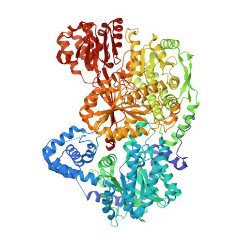
    Aldehyde dehydrogenases (ALDHs) catalyze the NAD(P) + -dependent oxidation of aldehydes to carboxylic acids and are important for metabolism and detoxification. Although the ALDH superfamily fold is well established, some ALDHs contain an uncharacterized domain of unknown function (DUF) near the C terminus of the polypeptide chain. Herein, we report the first structure of a protein containing the ALDH superfamily DUF. Proline utilization A from Sinorhizobium meliloti (SmPutA) is a 1233-residue bifunctional enzyme that contains the DUF in addition to proline dehydrogenase and l-glutamate-γ-semialdehyde dehydrogenase catalytic modules. Structures of SmPutA with a proline analog bound to the proline dehydrogenase site and NAD + bound to the ALDH site were determined in two space groups at 1.7-1.9 Å resolution. The DUF consists of a Rossmann dinucleotide-binding fold fused to a three-stranded β-flap. The Rossmann domain resembles the classic ALDH superfamily NAD + -binding domain, whereas the flap is strikingly similar to the ALDH superfamily dimerization domain. Paradoxically, neither structural element performs its implied function. Electron density maps show that NAD + does not bind to the DUF Rossmann fold, and small-angle X-ray scattering reveals a novel dimer that has never been seen in the ALDH superfamily. The structure suggests that the DUF is an adapter domain that stabilizes the aldehyde substrate binding loop and seals the substrate-channeling tunnel via tertiary structural interactions that mimic the quaternary structural interactions found in non-DUF PutAs. Kinetic data for SmPutA indicate a substrate-channeling mechanism, in agreement with previous studies of other PutAs.


  • Organizational Affiliation

    From the Departments of Chemistry and.


Macromolecules
Find similar proteins by:  (by identity cutoff)  |  3D Structure
Entity ID: 1
MoleculeChains Sequence LengthOrganismDetailsImage
Bifunctional protein PutA
A, B
1,235Sinorhizobium meliloti SM11Mutation(s): 0 
Gene Names: putASM11_chr0102
UniProt
Find proteins for F7X6I3 (Sinorhizobium meliloti (strain SM11))
Explore F7X6I3 
Go to UniProtKB:  F7X6I3
Entity Groups  
Sequence Clusters30% Identity50% Identity70% Identity90% Identity95% Identity100% Identity
UniProt GroupF7X6I3
Sequence Annotations
Expand
  • Reference Sequence
Small Molecules
Ligands 6 Unique
IDChains Name / Formula / InChI Key2D Diagram3D Interactions
FAD
Query on FAD

Download Ideal Coordinates CCD File 
C [auth A],
J [auth B]
FLAVIN-ADENINE DINUCLEOTIDE
C27 H33 N9 O15 P2
VWWQXMAJTJZDQX-UYBVJOGSSA-N
NAD
Query on NAD

Download Ideal Coordinates CCD File 
D [auth A],
K [auth B]
NICOTINAMIDE-ADENINE-DINUCLEOTIDE
C21 H27 N7 O14 P2
BAWFJGJZGIEFAR-NNYOXOHSSA-N
PG4
Query on PG4

Download Ideal Coordinates CCD File 
S [auth B]TETRAETHYLENE GLYCOL
C8 H18 O5
UWHCKJMYHZGTIT-UHFFFAOYSA-N
PGE
Query on PGE

Download Ideal Coordinates CCD File 
G [auth A]
H [auth A]
I [auth A]
N [auth B]
O [auth B]
G [auth A],
H [auth A],
I [auth A],
N [auth B],
O [auth B],
P [auth B],
Q [auth B],
R [auth B]
TRIETHYLENE GLYCOL
C6 H14 O4
ZIBGPFATKBEMQZ-UHFFFAOYSA-N
TFB
Query on TFB

Download Ideal Coordinates CCD File 
E [auth A],
L [auth B]
TETRAHYDROFURAN-2-CARBOXYLIC ACID
C5 H8 O3
UJJLJRQIPMGXEZ-BYPYZUCNSA-N
MG
Query on MG

Download Ideal Coordinates CCD File 
F [auth A],
M [auth B]
MAGNESIUM ION
Mg
JLVVSXFLKOJNIY-UHFFFAOYSA-N
Experimental Data & Validation

Experimental Data

  • Method: X-RAY DIFFRACTION
  • Resolution: 1.70 Å
  • R-Value Free: 0.222 
  • R-Value Work: 0.190 
  • R-Value Observed: 0.192 
  • Space Group: P 1 21 1
Unit Cell:
Length ( Å )Angle ( ˚ )
a = 101.4α = 90
b = 102.332β = 106.53
c = 125.909γ = 90
Software Package:
Software NamePurpose
Aimlessdata scaling
PHENIXrefinement
PDB_EXTRACTdata extraction
XDSdata reduction
PHASERphasing

Structure Validation

View Full Validation Report


Ligand Structure Quality Assessment 


Entry History & Funding Information

Deposition Data

  • Released Date: 2016-10-05 
  • Deposition Author(s): Tanner, J.J.

Funding OrganizationLocationGrant Number
National Institutes of Health/National Institute of General Medical Sciences (NIH/NIGMS)United StatesR01GM065546
National Institutes of Health/National Institute of General Medical Sciences (NIH/NIGMS)United StatesR01GM061068
National Institutes of Health/National Institute of General Medical Sciences (NIH/NIGMS)United StatesP30GM103335
National Science Foundation (NSF, United States)United StatesDBI-1156692

Revision History  (Full details and data files)

  • Version 1.0: 2016-10-05
    Type: Initial release
  • Version 1.1: 2016-10-19
    Changes: Database references
  • Version 1.2: 2016-11-23
    Changes: Database references
  • Version 1.3: 2017-09-20
    Changes: Author supporting evidence, Database references
  • Version 1.4: 2017-11-01
    Changes: Author supporting evidence
  • Version 1.5: 2019-11-27
    Changes: Author supporting evidence
  • Version 1.6: 2023-09-27
    Changes: Data collection, Database references, Derived calculations, Refinement description