


Crystal structure of truncated hemolysin A from P. mirabilis at 2.0 Angstroms in high salt
Novak, W.R.P., Bhattacharyya, B., Grilley, D.P., Weaver, T.M.To be published.
Experimental Data Snapshot
Starting Model: experimental
View more details
wwPDB Validation 3D Report Full Report

Entity ID: 1 | |||||
|---|---|---|---|---|---|
| Molecule | Chains | Sequence Length | Organism | Details | Image |
| Hemolysin | 242 | Proteus mirabilis | Mutation(s): 0 Gene Names: hpmA | 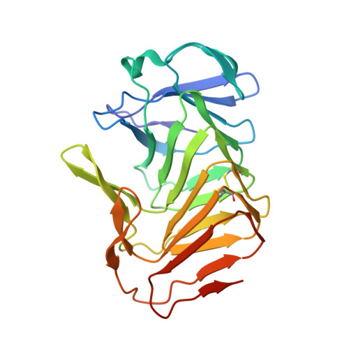 | |
UniProt | |||||
Entity Groups | |||||
| Sequence Clusters | 30% Identity50% Identity70% Identity90% Identity95% Identity100% Identity | ||||
| UniProt Group | P16466 | ||||
Sequence AnnotationsExpand | |||||
Reference Sequence | |||||
| Length ( Å ) | Angle ( ˚ ) |
|---|---|
| a = 34.44 | α = 90 |
| b = 80.15 | β = 90 |
| c = 96.61 | γ = 90 |
| Software Name | Purpose |
|---|---|
| MOSFLM | data reduction |
| Aimless | data scaling |
| MOLREP | phasing |
| PHENIX | refinement |
| PDB_EXTRACT | data extraction |


