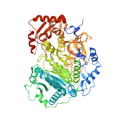
Crystal structure of Acetyl-CoA Synthetase in complex with ATP and Acetyl-AMP from Cryptococcus neoformans H99
Seattle Structural Genomics Center for Infectious Disease (SSGCID), Fox III, D., Delker, S.L., Potts, K.T., Numa, M.M., Edwards, T.E., Lorimer, D.D., Mutz, M.W., Krysan, D.J.To be published.