5K8A | pdb_00005k8a

NIST FAB


Experimental Data Snapshot

  • Method: X-RAY DIFFRACTION
  • Resolution: 2.00 Å
  • R-Value Free: 
    0.189 (Depositor), 0.225 (DCC) 
  • R-Value Work: 
    0.164 (Depositor), 0.203 (DCC) 
  • R-Value Observed: 
    0.166 (Depositor) 

Starting Model: experimental
View more details

wwPDB Validation 3D Report Full Report

Validation slider image for 5K8A

This is version 2.1 of the entry. See complete history

Literature

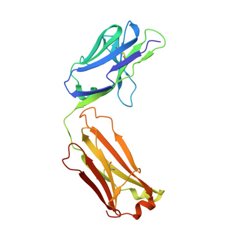
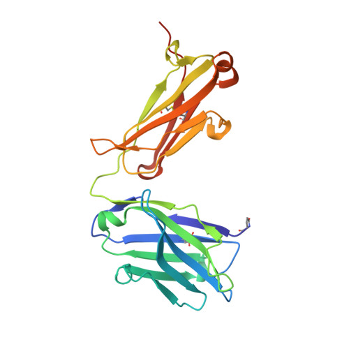
Data on crystal organization in the structure of the Fab fragment from the NIST reference antibody, RM 8671.

Gallagher, D.T.Karageorgos, I.Hudgens, J.W.Galvin, C.V.

(2018) Data Brief 16: 29-36

  • DOI: https://doi.org/10.1016/j.dib.2017.11.013
  • Primary Citation Related Structures: 
    5K8A

  • PubMed Abstract: 

    The reported data describe the crystallization, crystal packing, structure determination and twinning of the unliganded Fab (antigen-binding fragment) from the NISTmAb (standard reference material 8671). The raw atomic coordinates are available as Protein Data Bank structure 5K8A and biological aspects are described in the article, (Karageorgos et al., 2017) [1]. Crystal data show that the packing is unique, and show the basis for the crystal's twinned growth. Twinning is a common and often serious problem in protein structure determination by x-ray crystallography [2]. In the present case the twinning is due to a small deviation (about 0.3 nm) from 4-fold symmetry in the primary intermolecular interface. The deviation produces pseudosymmetry, generating slightly different conformations of the protein, and alternating strong and weak forms of key packing interfaces throughout the lattice.


  • Organizational Affiliation
    • Biomolecular Measurement Division, National Institute of Standards & Technology, Rockville, MD 20850, United States.

Macromolecule Content 

  • Total Structure Weight: 190.74 kDa 
  • Atom Count: 13,629 
  • Modeled Residue Count: 1,744 
  • Deposited Residue Count: 1,760 
  • Unique protein chains: 2

Macromolecules

Find similar proteins by:|  3D Structure
Entity ID: 1
MoleculeChains  Sequence LengthOrganismDetailsImage
humanized Fab Lite chainA [auth L],
C [auth M],
E [auth A],
G [auth E]
213Mus musculusMutation(s): 0 
Find similar proteins by:|  3D Structure
Entity ID: 2
MoleculeChains  Sequence LengthOrganismDetailsImage
Heavy chainB [auth H],
D [auth V],
F [auth B],
H [auth F]
227Mus musculusMutation(s): 0 

Small Molecules

Modified Residues  1 Unique
IDChains TypeFormula2D DiagramParent
PCA
Query on PCA
B [auth H],
D [auth V],
F [auth B],
H [auth F]
L-PEPTIDE LINKINGC5 H7 N O3GLN

Experimental Data & Validation

Experimental Data

  • Method: X-RAY DIFFRACTION
  • Resolution: 2.00 Å
  • R-Value Free:  0.189 (Depositor), 0.225 (DCC) 
  • R-Value Work:  0.164 (Depositor), 0.203 (DCC) 
  • R-Value Observed: 0.166 (Depositor) 
Space Group: I 2 2 2
Unit Cell:
Length ( Å )Angle ( ˚ )
a = 149.156α = 90
b = 149.203β = 90
c = 195.032γ = 90
Software Package:
Software NamePurpose
HKL-2000data collection
HKL-2000data scaling
PHASERphasing
REFMACrefinement
PDB_EXTRACTdata extraction
HKL-2000data reduction

Structure Validation

View Full Validation Report


Entry History 

Deposition Data

Revision History  (Full details and data files)

  • Version 1.0: 2017-08-09
    Type: Initial release
  • Version 1.1: 2019-01-23
    Changes: Data collection, Database references
  • Version 2.0: 2023-09-27
    Changes: Data collection, Database references, Polymer sequence, Refinement description
  • Version 2.1: 2024-10-23
    Changes: Structure summary