5K10 | pdb_00005k10

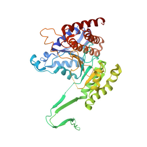
Cryo-EM structure of isocitrate dehydrogenase (IDH1)


Experimental Data Snapshot

  • Method: ELECTRON MICROSCOPY
  • Resolution: 3.80 Å
  • Aggregation State: PARTICLE 
  • Reconstruction Method: SINGLE PARTICLE 

Starting Model: experimental
View more details

wwPDB Validation 3D Report Full Report

Validation slider image for 5K10

This is version 1.4 of the entry. See complete history. 

Literature

Breaking Cryo-EM Resolution Barriers to Facilitate Drug Discovery.

Merk, A., Bartesaghi, A., Banerjee, S., Falconieri, V., Rao, P., Davis, M.I., Pragani, R., Boxer, M.B., Earl, L.A., Milne, J.L., Subramaniam, S.

(2016) Cell 165: 1698-1707

  • DOI: https://doi.org/10.1016/j.cell.2016.05.040
  • Primary Citation Related Structures: 
    5K0Z, 5K10, 5K11, 5K12

  • PubMed Abstract: 

    Recent advances in single-particle cryoelecton microscopy (cryo-EM) are enabling generation of numerous near-atomic resolution structures for well-ordered protein complexes with sizes ≥ ∼200 kDa. Whether cryo-EM methods are equally useful for high-resolution structural analysis of smaller, dynamic protein complexes such as those involved in cellular metabolism remains an important question. Here, we present 3.8 Å resolution cryo-EM structures of the cancer target isocitrate dehydrogenase (93 kDa) and identify the nature of conformational changes induced by binding of the allosteric small-molecule inhibitor ML309. We also report 2.8-Å- and 1.8-Å-resolution structures of lactate dehydrogenase (145 kDa) and glutamate dehydrogenase (334 kDa), respectively. With these results, two perceived barriers in single-particle cryo-EM are overcome: (1) crossing 2 Å resolution and (2) obtaining structures of proteins with sizes < 100 kDa, demonstrating that cryo-EM can be used to investigate a broad spectrum of drug-target interactions and dynamic conformational states.


  • Organizational Affiliation: 
    • Laboratory of Cell Biology, Center for Cancer Research, National Cancer Institute, NIH, Bethesda, MD 20892, USA.

Macromolecule Content 

  • Total Structure Weight: 94.16 kDa 
  • Atom Count: 6,338 
  • Modeled Residue Count: 794 
  • Deposited Residue Count: 822 
  • Unique protein chains: 1

Macromolecules

Find similar proteins by:|  3D Structure
Entity ID: 1
MoleculeChains  Sequence LengthOrganismDetailsImage
Isocitrate dehydrogenase [NADP] cytoplasmic
A, B
411Homo sapiensMutation(s): 0 
Gene Names: IDH1, PICD
EC: 1.1.1.42
UniProt & NIH Common Fund Data Resources
Find proteins for O75874 (Homo sapiens)
Explore O75874 
Go to UniProtKB:  O75874
PHAROS:  O75874
GTEx:  ENSG00000138413 
Entity Groups
Sequence Clusters30% Identity50% Identity70% Identity90% Identity95% Identity100% Identity
UniProt GroupO75874
Sequence Annotations
Expand
Reference Sequence

Small Molecules

Ligands 1 Unique
IDChains Name / Formula / InChI Key2D Diagram3D Interactions
NDP

Query on NDP


Download:Ideal Coordinates CCD File
C [auth A],
D [auth B]
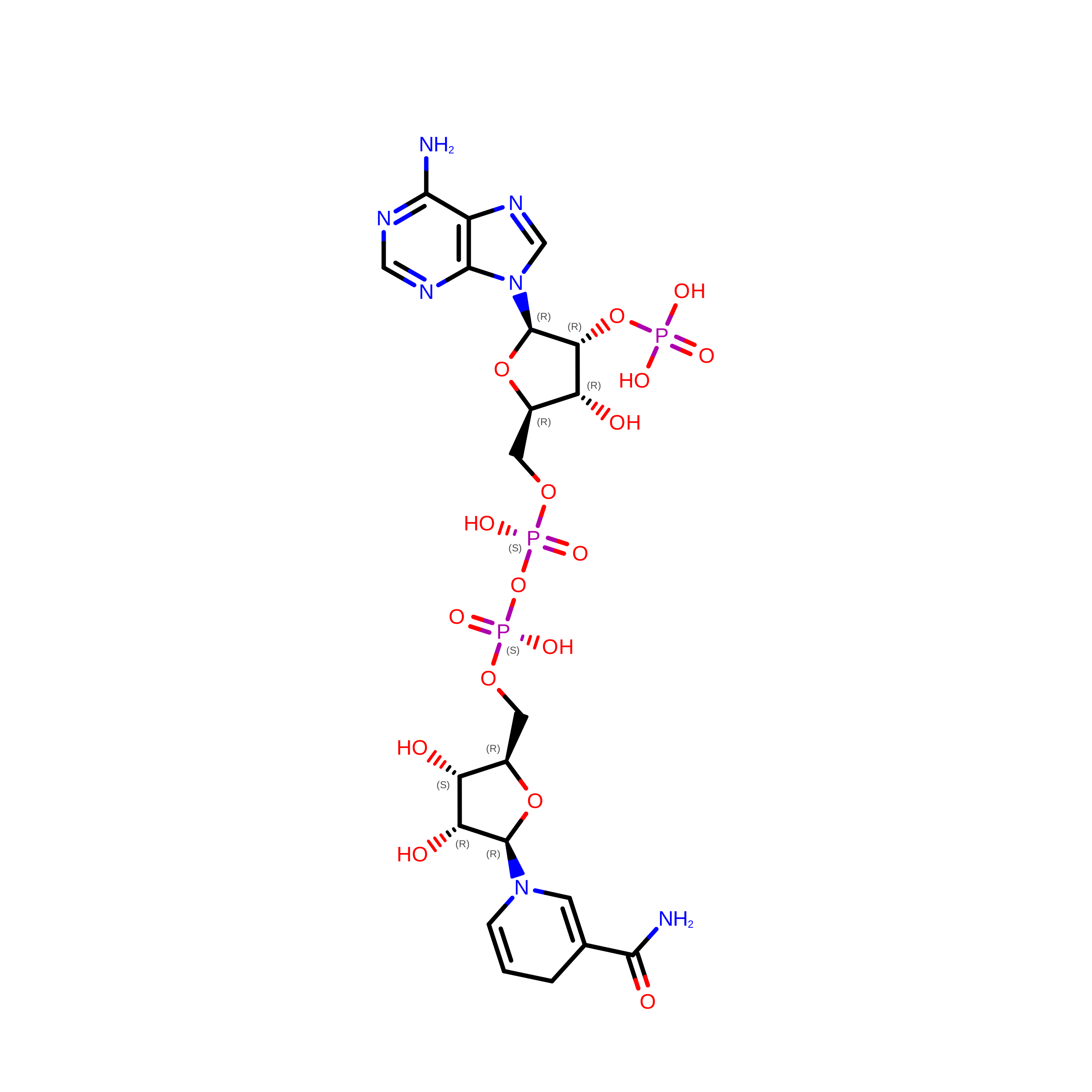
NADPH DIHYDRO-NICOTINAMIDE-ADENINE-DINUCLEOTIDE PHOSPHATE
C21 H30 N7 O17 P3
ACFIXJIJDZMPPO-NNYOXOHSSA-N

Experimental Data & Validation

Experimental Data

  • Method: ELECTRON MICROSCOPY
  • Resolution: 3.80 Å
  • Aggregation State: PARTICLE 
  • Reconstruction Method: SINGLE PARTICLE 
EM Software:
TaskSoftware PackageVersion
MODEL REFINEMENTPHENIX
MODEL REFINEMENTCoot
RECONSTRUCTIONFREALIGN9.10

Structure Validation

View Full Validation Report


Entry History 

Deposition Data

Revision History  (Full details and data files)

  • Version 1.0: 2016-06-08
    Type: Initial release
  • Version 1.1: 2016-06-29
    Changes: Database references
  • Version 1.2: 2016-11-23
    Changes: Other
  • Version 1.3: 2018-07-18
    Changes: Data collection, Experimental preparation
  • Version 1.4: 2024-03-06
    Changes: Data collection, Database references, Refinement description