5K0P

Crystal structure of the archaeosine synthase QueF-Like in the apo form


Experimental Data Snapshot

  • Method: X-RAY DIFFRACTION
  • Resolution: 1.94 Å
  • R-Value Free: 0.238 
  • R-Value Work: 0.155 
  • R-Value Observed: 0.159 

wwPDB Validation   3D Report Full Report


This is version 1.4 of the entry. See complete history


Literature

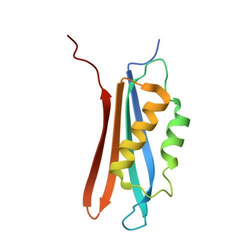
Crystal structure of the archaeosine synthase QueF-like-Insights into amidino transfer and tRNA recognition by the tunnel fold.

Mei, X.Alvarez, J.Bon Ramos, A.Samanta, U.Iwata-Reuyl, D.Swairjo, M.A.

(2017) Proteins 85: 103-116

  • DOI: https://doi.org/10.1002/prot.25202
  • Primary Citation of Related Structures:  
    5JYX, 5K0P

  • PubMed Abstract: 

    The tunneling-fold (T-fold) structural superfamily has emerged as a versatile protein scaffold of diverse catalytic activities. This is especially evident in the pathways to the 7-deazaguanosine modified nucleosides of tRNA queuosine and archaeosine. Four members of the T-fold superfamily have been confirmed in these pathways and here we report the crystal structure of a fifth enzyme; the recently discovered amidinotransferase QueF-Like (QueF-L), responsible for the final step in the biosynthesis of archaeosine in the D-loop of tRNA in a subset of Crenarchaeota. QueF-L catalyzes the conversion of the nitrile group of the 7-cyano-7-deazaguanine (preQ 0 ) base of preQ 0 -modified tRNA to a formamidino group. The structure, determined in the presence of preQ 0 , reveals a symmetric T-fold homodecamer of two head-to-head facing pentameric subunits, with 10 active sites at the inter-monomer interfaces. Bound preQ 0 forms a stable covalent thioimide bond with a conserved active site cysteine similar to the intermediate previously observed in the nitrile reductase QueF. Despite distinct catalytic functions, phylogenetic distributions, and only 19% sequence identity, the two enzymes share a common preQ 0 binding pocket, and likely a common mechanism of thioimide formation. However, due to tight twisting of its decamer, QueF-L lacks the NADPH binding site present in QueF. A large positively charged molecular surface and a docking model suggest simultaneous binding of multiple tRNA molecules and structure-specific recognition of the D-loop by a surface groove. The structure sheds light on the mechanism of nitrile amidation, and the evolution of diverse chemistries in a common fold. Proteins 2016; 85:103-116. © 2016 Wiley Periodicals, Inc.


  • Organizational Affiliation

    Department of Chemistry and Biochemistry, San Diego State University- 5500 Campanile Drive, San Diego, California, 92182.


Macromolecules
Find similar proteins by:  (by identity cutoff)  |  3D Structure
Entity ID: 1
MoleculeChains Sequence LengthOrganismDetailsImage
Archeaosine synthase QueF-Like
A, B, C, D, E
A, B, C, D, E, F, G, H, I, J
109Pyrobaculum calidifontis JCM 11548Mutation(s): 0 
Gene Names: Pcal_0221
UniProt
Find proteins for A3MSP1 (Pyrobaculum calidifontis (strain DSM 21063 / JCM 11548 / VA1))
Explore A3MSP1 
Go to UniProtKB:  A3MSP1
Entity Groups  
Sequence Clusters30% Identity50% Identity70% Identity90% Identity95% Identity100% Identity
UniProt GroupA3MSP1
Sequence Annotations
Expand
  • Reference Sequence
Small Molecules
Ligands 3 Unique
IDChains Name / Formula / InChI Key2D Diagram3D Interactions
PG4
Query on PG4

Download Ideal Coordinates CCD File 
V [auth I]TETRAETHYLENE GLYCOL
C8 H18 O5
UWHCKJMYHZGTIT-UHFFFAOYSA-N
PGE
Query on PGE

Download Ideal Coordinates CCD File 
M [auth C],
O [auth D],
R [auth F]
TRIETHYLENE GLYCOL
C6 H14 O4
ZIBGPFATKBEMQZ-UHFFFAOYSA-N
SCN
Query on SCN

Download Ideal Coordinates CCD File 
K [auth A]
L [auth B]
N [auth C]
P [auth D]
Q [auth E]
K [auth A],
L [auth B],
N [auth C],
P [auth D],
Q [auth E],
S [auth F],
T [auth G],
U [auth H],
W [auth I],
X [auth J]
THIOCYANATE ION
C N S
ZMZDMBWJUHKJPS-UHFFFAOYSA-M
Experimental Data & Validation

Experimental Data

  • Method: X-RAY DIFFRACTION
  • Resolution: 1.94 Å
  • R-Value Free: 0.238 
  • R-Value Work: 0.155 
  • R-Value Observed: 0.159 
  • Space Group: P 1 21 1
Unit Cell:
Length ( Å )Angle ( ˚ )
a = 62.21α = 90
b = 124.995β = 112.27
c = 74.404γ = 90
Software Package:
Software NamePurpose
REFMACrefinement
HKL-2000data scaling
PDB_EXTRACTdata extraction
HKL-2000data reduction
PHASERphasing

Structure Validation

View Full Validation Report


Entry History & Funding Information

Deposition Data


Funding OrganizationLocationGrant Number
National Science Foundation (NSF, United States)United StatesCHE-1309323

Revision History  (Full details and data files)

  • Version 1.0: 2016-11-09
    Type: Initial release
  • Version 1.1: 2017-09-13
    Changes: Author supporting evidence, Derived calculations
  • Version 1.2: 2019-07-10
    Changes: Data collection, Database references
  • Version 1.3: 2019-11-27
    Changes: Author supporting evidence
  • Version 1.4: 2023-09-27
    Changes: Data collection, Database references, Refinement description