5K0H | pdb_00005k0h

Human factor Xa in complex with synthetic inhibitor benzylsulfonyl-dSer(Benzyl)-Gly-4-amidinobenzylamide


Experimental Data Snapshot

  • Method: X-RAY DIFFRACTION
  • Resolution: 2.20 Å
  • R-Value Free: 
    0.261 (Depositor) 
  • R-Value Work: 
    0.214 (Depositor) 
  • R-Value Observed: 
    0.214 (Depositor) 

Starting Model: experimental
View more details

wwPDB Validation 3D Report Full Report

Validation slider image for 5K0H

This is version 1.2 of the entry. See complete history

Literature

New substrate analogue inhibitors of factor Xa containing 4-amidinobenzylamide as P1 residue: part 1.

Schweinitz, A.Sturzebecher, A.Sturzebecher, U.Schuster, O.Sturzebecher, J.Steinmetzer, T.

(2006) Med Chem 2: 349-361

  • DOI: https://doi.org/10.2174/157340606777724040
  • Primary Citation Related Structures: 
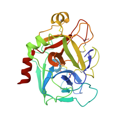
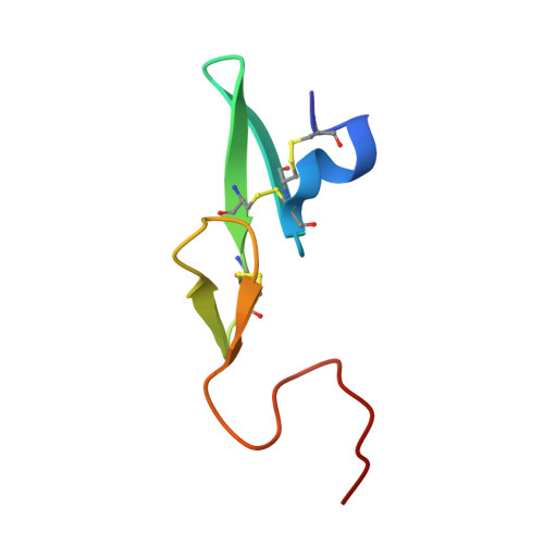
    5K0H

  • PubMed Abstract: 

    The trypsin-like serine protease factor Xa (fXa) is located at the convergence point of the intrinsic and extrinsic coagulation cascade, and therefore has emerged as an attractive target for the design of novel anticoagulants. During the development of substrate-analogue urokinase inhibitors we have found that the protection of the P3-dSer side chain leads to a scaffold of potent fXa inhibitors with the general structure R1-SO2-dSer(R2)-Gly-4-amidinobenzylamide. The first lead (3) with an N-terminal benzylsulfonyl group and dSer(tBu) as P3 residue inhibits human fXa with a Ki of 14 nM. A variety of derivatives with modified P4, P3, and P2 residues have been investigated in terms of inhibition of fXa and related proteases and for their anticoagulant potency and elimination behaviour. Most inhibitors were rapidly cleared from the circulation of rats. However, compound 48 (Ki= 3.5 nM), one of the most potent and selective inhibitors containing a dArg as P3 residue was relatively slowly eliminated (t1/2 approximately 1 h). Inhibitor 48 doubled clotting times in human plasma at 0.32 microM (aPTT) and 0.28 microM (PT), and is approximately 10-fold more potent than the reference fXa inhibitor DX-9065a in the inhibition of the prothrombinase complex. The structures of two inhibitors in complex with human fXa were solved by X-ray crystallography.


  • Organizational Affiliation
    • Curacyte Chemistry GmbH, Winzerlaer Str. 2, D-07745 Jena, Germany.

Macromolecule Content 

  • Total Structure Weight: 32.52 kDa 
  • Atom Count: 2,412 
  • Modeled Residue Count: 285 
  • Deposited Residue Count: 285 
  • Unique protein chains: 2

Macromolecules

Find similar proteins by:|  3D Structure
Entity ID: 1
MoleculeChains  Sequence LengthOrganismDetailsImage
Coagulation factor X234Homo sapiensMutation(s): 0 
EC: 3.4.21.6
UniProt & NIH Common Fund Data Resources
Find proteins for P00742 (Homo sapiens)
Explore P00742 
Go to UniProtKB:  P00742
PHAROS:  P00742
GTEx:  ENSG00000126218 
Entity Groups
Sequence Clusters30% Identity50% Identity70% Identity90% Identity95% Identity100% Identity
UniProt GroupP00742
Sequence Annotations
Expand
Reference Sequence
Find similar proteins by:|  3D Structure
Entity ID: 2
MoleculeChains  Sequence LengthOrganismDetailsImage
Coagulation factor X51Homo sapiensMutation(s): 0 
EC: 3.4.21.6
UniProt & NIH Common Fund Data Resources
Find proteins for P00742 (Homo sapiens)
Explore P00742 
Go to UniProtKB:  P00742
PHAROS:  P00742
GTEx:  ENSG00000126218 
Entity Groups
Sequence Clusters30% Identity50% Identity70% Identity90% Identity95% Identity100% Identity
UniProt GroupP00742
Sequence Annotations
Expand
Reference Sequence

Small Molecules

Ligands 2 Unique
IDChains Name / Formula / InChI Key2D Diagram3D Interactions
6PK

Query on 6PK


Download:Ideal Coordinates CCD File
E [auth A]O-benzyl-N-(benzylsulfonyl)-D-seryl-N-[(4-carbamimidoylphenyl)methyl]glycinamide
C27 H31 N5 O5 S
RXJPCNAPBKYKMA-XMMPIXPASA-N
CA

Query on CA


Download:Ideal Coordinates CCD File
C [auth A],
D [auth A]
CALCIUM ION
Ca
BHPQYMZQTOCNFJ-UHFFFAOYSA-N
Binding Affinity Annotations 
IDSourceBinding Affinity
6PK BindingDB:  5K0H Ki: 29 (nM) from 1 assay(s)

Experimental Data & Validation

Experimental Data

  • Method: X-RAY DIFFRACTION
  • Resolution: 2.20 Å
  • R-Value Free:  0.261 (Depositor) 
  • R-Value Work:  0.214 (Depositor) 
  • R-Value Observed: 0.214 (Depositor) 
Space Group: P 21 21 21
Unit Cell:
Length ( Å )Angle ( ˚ )
a = 56.28α = 90
b = 72.4β = 90
c = 77.13γ = 90
Software Package:
Software NamePurpose
CNXrefinement
Omodel building

Structure Validation

View Full Validation Report


Entry History 

Deposition Data

Revision History  (Full details and data files)

  • Version 1.0: 2016-07-06
    Type: Initial release
  • Version 1.1: 2024-01-10
    Changes: Data collection, Database references, Refinement description
  • Version 1.2: 2024-11-06
    Changes: Structure summary