Crystal Structures of Group B Streptococcus Glyceraldehyde-3-Phosphate Dehydrogenase: Apo-Form, Binary and Ternary Complexes.
Schormann, N., Ayres, C.A., Fry, A., Green, T.J., Banerjee, S., Ulett, G.C., Chattopadhyay, D.(2016) PLoS One 11: e0165917-e0165917
- PubMed: 27875551 Search on PubMedSearch on PubMed Central
- DOI: https://doi.org/10.1371/journal.pone.0165917
- Primary Citation Related Structures:
5JY6, 5JYA, 5JYE, 5JYF - PubMed Abstract:
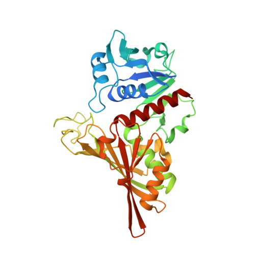
Glyceraldehyde 3-phosphate dehydrogenase or GAPDH is an evolutionarily conserved glycolytic enzyme. It catalyzes the two step oxidative phosphorylation of D-glyceraldehyde 3-phosphate into 1,3-bisphosphoglycerate using inorganic phosphate and NAD+ as cofactor. GAPDH of Group B Streptococcus is a major virulence factor and a potential vaccine candidate. Moreover, since GAPDH activity is essential for bacterial growth it may serve as a possible drug target. Crystal structures of Group B Streptococcus GAPDH in the apo-form, two different binary complexes and the ternary complex are described here. The two binary complexes contained NAD+ bound to 2 (mixed-holo) or 4 (holo) subunits of the tetrameric protein. The structure of the mixed-holo complex reveals the effects of NAD+ binding on the conformation of the protein. In the ternary complex, the phosphate group of the substrate was bound to the new Pi site in all four subunits. Comparison with the structure of human GAPDH showed several differences near the adenosyl binding pocket in Group B Streptococcus GAPDH. The structures also reveal at least three surface-exposed areas that differ in amino acid sequence compared to the corresponding areas of human GAPDH.
- Department of Medicine, University of Alabama at Birmingham, Birmingham, Alabama 35294, United States of America.
Organizational Affiliation: